. 2007 Sep;194(1):125-33.
doi: 10.1016/j.atherosclerosis.2006.10.013. Epub 2006 Nov 15.

Angiotensin II induces IL-6 expression and the Jak-STAT3 pathway in aortic adventitia of LDL receptor-deficient mice

Affiliations

Angiotensin II induces IL-6 expression and the Jak-STAT3 pathway in aortic adventitia of LDL receptor-deficient mice

Adrian Recinos 3rd et al. Atherosclerosis. 2007 Sep.

Abstract

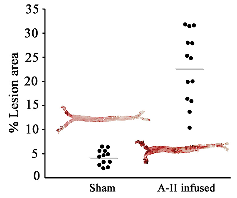
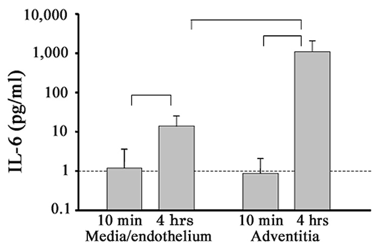
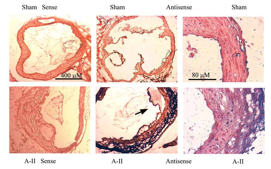
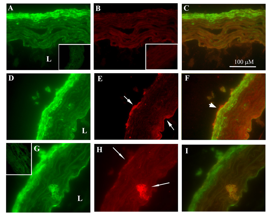
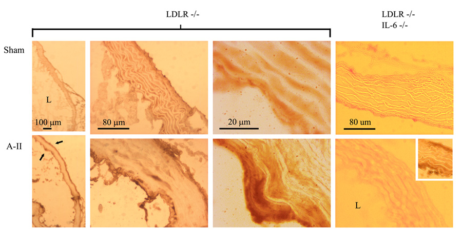
Angiotensin II (A-II), the major effector peptide of the renin angiotensin system potently accelerates progression of atherosclerosis. To investigate its effects on vascular inflammatory mechanisms, we elucidated vascular cytokine expression during early lesion development in A-II-infused atherosclerosis-prone LDLR-/- mice. Male LDLR-/- mice were placed on a "Western" high-fat diet for 4 weeks, followed by sham or A-II infusion for 7 weeks. Equal blood pressures and elevations in serum lipids were seen in both groups. Mice were sacrificed when significant A-II-induced plaque development was first detectable, aortae were explanted and culture media assayed for secreted cytokines. Nine cytokines were significantly induced with interleukin-6 (IL-6) being the most highly secreted. Local IL-6 production was confirmed by in situ mRNA hybridization and immunostaining, where the most abundant IL-6 was found in the aortic adventitia, with lesser production by the medial and intimal layers. Immunofluorescence colocalization showed IL-6 expression by fibroblasts and activated macrophages. Activation of downstream IL-6 signaling mediated by the Jak-STAT3 pathway was demonstrated by inducible phospho-Tyr705-STAT3 formation in the adventitia and endothelium (of IL-6+/+ mice only). These findings define cytokine profiles in the A-II infusion model and demonstrate that IL-6, produced by activated macrophages and fibroblasts in the adventitia, induces the Jak-STAT3 pathway during early A-II-induced atherosclerosis.


Figures

Figure 1
Figure 1
En face histomorphometry of atherosclerotic lesions. At time of sacrifice, aortae were harvested, dissected en face from the arch to just beyond the iliac bifurcation and stained with oil red O. Dot plots indicate percent aortic lesion areas for surviving individual mice by infusion group [means and (SD), sham 4.3 (1.6), A-II 22.9 (7.3), P<10−6, Student’s t-test]. Insets: photographs of 2 representative aortae, sham- and AII-infused, 4.4 and 25.3% lesion areas, respectively.
Figure 2
Figure 2
A-II-induced aortic IL-6 secretion, adventitial versus medial/endothelial layers. Separately explanted aortic adventitial and medial/endothelial regions were assayed for IL-6 secretion after 10 min and after 4 h of incubation (shown are means ± SD). Indicated are comparisons of secretion increases with time, and secretion increase in adventitia versus media/endothelium at 4 h (n=5, all P<0.05, ANOVA).
Figure 3
Figure 3
In Situ hybridization of IL-6 mRNA in aortic cross sections. Aortic cross sections were hybridized with sense (control) and anti-sense IL-6 cRNA probes. Upper panels, Aortae from sham-infused mice probed with sense (far left) and anti-sense IL-6 probes. Two different magnifications are shown (40X and 200X). Lower panels, aortae from A-II-infused mice were hybridized as in the upper panels. Predominant IL-6 staining is in the adventitial layer (purple) with additional diffuse staining in the media and endothelium. Arrow: Endothelial IL-6 staining in region of lesion development.
Figure 4
Figure 4
Colocalization immunofluorescence histochemistry of IL-6 with fibroblasts and macrophages. Transverse cryosections (6 µm) of proximal aorta from sham- (A–C) and A-II-infused (D–I) mice were FITC-stained green for fibroblasts (A and D), FITC-stained for activated macrophages (G) or Texas Red-stained (B,E and H) for IL-6. In each row, the 3rd column is the merged image (yellow indicates colocalization). L, lumen. With A-II infusion, IL-6 is predominantly detected in adventitia (E and H, upper arrow), but also in endothelial (E, lower arrow) and medial areas (H, lower arrow). IL-6 colocalizing with fibroblasts (F, thick arrow) and with activated macrophages (I) are indicated. Insets: Control immunofluorescence using FITC- or Texas Red-conjugated secondary Ab only on sections from sham-treated animals (similar A-II-treated secondary Ab controls were indistinguishable from those shown). (X 600).
Figure 5
Figure 5
Phospho-Tyr705 STAT3 accumulation induced by A-II infusion. In situ histochemistry for phospho-Tyr705 STAT3 was performed in aortic cryosections of LDLR−/− and LDLR−/−/IL-6−/− mice. L, lumen (bottom left in all photos). Top panels, sham-infused vessels show little accumulation of phospho-STAT3 (dark brown staining). Bottom panels, A-II-infused aortae show significant staining in both adventitial and endothelial layers (arrows), except in the double-knock-out mouse genotype (right hand panels). Inset: Aorta from LDLR−/−mouse stained in parallel with double-knock-outs (positive control). Original microscopy at 40X, 200X and 1000X.

Similar articles

Cited by

References

    1. Daugherty A, Cassis L. Angiotensin II-mediated development of vascular diseases. Trends in Cardiovasc Med. 2004;14:117–120. - PubMed
    1. Kon V, Jabs K. Angiotensin in atherosclerosis. Curr Opin Nephrol Hypertens. 2004;13:291–297. - PubMed
    1. Brasier AR, Recinos A, Eledrisi MS. Vascular inflammation and the renin-angiotensin system. Arterioscler Thromb Vasc Biol. 2002;22:1257–1266. - PubMed
    1. Brasier AR, Jamaluddin M, Han Y, Patterson C, Runge MS. Angiotensin II induces gene transcription through cell-type-dependent effects on the nuclear factor-kappaB (NF-kappaB)transcription factor. Mol Cell Biochem. 2000:155–699. - PubMed
    1. Murphy TJ, Alexander RW, Griendling KK, Runge MS, Bernstein KE. Isolation of a cDNA encoding the vascular type-1 angiotensin II receptor. Nature. 1991;351:233–236. - PubMed

Publication types

MeSH terms