. 2006 Nov 28;103(48):18160-5.
doi: 10.1073/pnas.0606508103. Epub 2006 Nov 16.

Fragile X mental retardation protein controls trailer hitch expression and cleavage furrow formation in Drosophila embryos

Affiliations

Fragile X mental retardation protein controls trailer hitch expression and cleavage furrow formation in Drosophila embryos

Kate Monzo et al. Proc Natl Acad Sci U S A. .

Abstract

During the cleavage stage of animal embryogenesis, cell numbers increase dramatically without growth, and a shift from maternal to zygotic genetic control occurs called the midblastula transition. Although these processes are fundamental to animal development, the molecular mechanisms controlling them are poorly understood. Here, we demonstrate that Drosophila fragile X mental retardation protein (dFMRP) is required for cleavage furrow formation and functions within dynamic cytoplasmic ribonucleoprotein (RNP) bodies during the midblastula transition. dFMRP is observed to colocalize with the cytoplasmic RNP body components Maternal expression at 31B (ME31B) and Trailer Hitch (TRAL) in a punctate pattern throughout the cytoplasm of cleavage-stage embryos. Complementary biochemistry demonstrates that dFMRP does not associate with polyribosomes, consistent with their reported exclusion from many cytoplasmic RNP bodies. By using a conditional mutation in small bristles (sbr), which encodes an mRNA nuclear export factor, to disrupt the normal cytoplasmic accumulation of zygotic transcripts at the midblastula transition, we observe the formation of giant dFMRP/TRAL-associated structures, suggesting that dFMRP and TRAL dynamically regulate RNA metabolism at the midblastula transition. Furthermore, we show that dFMRP associates with endogenous tral mRNA and is required for normal TRAL protein expression and localization, revealing it as a previously undescribed target of dFMRP control. We also show genetically that tral itself is required for cleavage furrow formation. Together, these data suggest that in cleavage-stage Drosophila embryos, dFMRP affects protein expression by controlling the availability and/or competency of specific transcripts to be translated.


Conflict of interest statement

The authors declare no conflict of interest.

Figures

Fig. 1.
Fig. 1.
Maternal expression of dfmr1 is required for cellularization. (a) Immunoblots of 15 μg of Oregon-R (WT) and fmr1 (fmr13/Df) adult female extracts probed for dFMRP and Myosin II (MYOII, control). (b) The number of F1 embryos laid (white plus gray) and hatched (gray) are shown as bars for each cross. Hatch percentages (hatched/laid) are indicated on each bar. The striped region of bar 4 indicates the number of fmr1/TM3-GFP, fmr1+ hatchlings. (c) The average rate of furrow ingression is shown for embryos derived from the indicated female genotypes at 25°C. fmr1 embryos with severe furrowing defects (1d Right) were not quantified. Error bars and n indicate the standard deviation (SD) and number of movies measured, respectively. (d) Sequential frames from representative differential interference contrast movies of WT and fmr1 embryos undergoing normal (Left), delayed (Center), and severely disrupted (Right) cellularization at 25°C. The percentage of fmr1 embryos in each phenotypic class is shown in parentheses. White arrowheads and brackets indicate the furrow front position and nuclear elongation, respectively. Times (t = minutes) are relative to nuclear cycle 14 onset. (Scale bar: 10 μm.)
Fig. 2.
Fig. 2.
dFMRP localizes to punctate cytoplasmic structures in cleavage-stage embryos. IF analysis of fixed WT embryos at progressive stages of cellularization shows punctate dFMRP localization throughout the cytoplasm (Left) and corresponding cortical F-actin marking plasma membrane furrows (Right). dFMRP puncta often are positioned at the furrow front (arrow and bracket). Asterisks indicate nuclei. (Scale bar: 10 μm.)
Fig. 3.
Fig. 3.
dFMRP associates with cytoplasmic RNP bodies in cleavage-stage embryos. (a) IF analysis of fixed WT cellularizing embryos shows considerable colocalization of dFMRP with TRAL and ME31B throughout the cytoplasm (apical cytoplasm shown) and partial colocalization with dAGO2 (only observed in the basal cytoplasm) in oblique optical sections. Arrows indicate examples of colocalization. (Scale bar: 10 μm.) (b) A UV absorbance trace (A254) indicates the positions of ribosomal subunits and polyribosomes across fractions 1–22 from a sucrose gradient. Immunoblots reveal the sedimentation profiles of proteins indicated at the left. (c and d) Immunoblots showing supernatant (sup) and pellet (pel) fractions from IPs performed with anti-FLAG (control) or anti-dFMRP antibodies (c) and BSA (control) or anti-TRAL antibody (d) by using WT embryo extracts. Blots were probed for the proteins indicated at the left. A weak nonspecific TRAL signal is observed in both pellets of c.
Fig. 4.
Fig. 4.
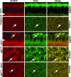
dFMRP/TRAL cytoplasmic RNP bodies are dramatically affected by disrupting the MBT. IF analysis of cellularizing WT or sbrts148 embryos fixed at 32°C or 20°C (indicated at left) shows dFMRP and TRAL localization in sagittal and surface views (indicated at right). The WT and sbrts148 embryos were shifted to 32°C at 5 and 15 min into nuclear cycle 14, respectively. Arrows indicate colocalization. Arrowheads indicate the furrow front. (Scale bars: 10 μm.)
Fig. 5.
Fig. 5.
tral mRNA is a target of dFMRP regulation. (a and b) IF analysis of fixed WT (Left) and fmr1 (Right) cellularizing embryos show Futsch (a Top), Lava Lamp (LVA) (a Middle), and F-actin (a Bottom) and TRAL (b Top and Center) and dFMRP (b Bottom) localization. Quantified fluorescence signal intensity (level in fmr1 / level in WT) is indicated in a Lower Right. (b) Sagittal (Top) and surface (Middle) optical sections show abnormal TRAL structures in the apical cytoplasm of fmr1 embryos. (Scale bar: 10 μm.) (c) Quantitative immunoblots show levels of the proteins indicated to the left in WT and fmr1 cellularizing embryo extracts. Each signal was independently normalized to an internal loading control Myosin II (MYOII). The ratios of normalized signal intensities (fmr1 / WT) are shown to the right (dAGO2 ratio = 0.85, data not shown). (d) Quantification of futsch, tral, and me31B mRNA levels in cellularizing WT and fmr1 embryos by real-time PCR shows a 2.7-fold increase in tral mRNA in fmr1 embryos. futsch and me31B mRNA levels show no significant difference. (e) Real-time PCR of RpL32 and tral mRNA in anti-dFMRP immunoprecipitates from cellularizing WT and fmr1 embryo extracts. Significance was assessed by using the Student's t test (∗∗, P ≤ 0.005); P = 0.003 in d; and P = 0.001 in e). Error bars indicate SDs.

References

    1. O'Donnell WT, Warren ST. Annu Rev Neurosci. 2002;25:315–338. - PubMed
    1. Laggerbauer B, Ostareck D, Keidel EM, Ostareck-Lederer A, Fischer U. Hum Mol Genet. 2001;10:329–338. - PubMed
    1. Li Z, Zhang Y, Ku L, Wilkinson KD, Warren ST, Feng Y. Nucleic Acids Res. 2001;29:2276–2283. - PMC - PubMed
    1. Zalfa F, Achsel T, Bagni C. Curr Opin Neurobiol. 2006;16:265–269. - PubMed
    1. Caudy AA, Myers M, Hannon GJ, Hammond SM. Genes Dev. 2002;16:2491–2496. - PMC - PubMed

Publication types

MeSH terms