. 2007 Feb;175(2):643-57.
doi: 10.1534/genetics.106.061929. Epub 2006 Nov 16.

Genes affecting cell competition in Drosophila

Affiliations

Genes affecting cell competition in Drosophila

David M Tyler et al. Genetics. 2007 Feb.

Abstract

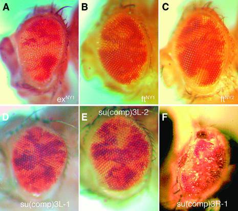
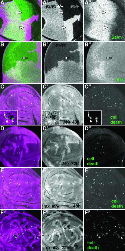
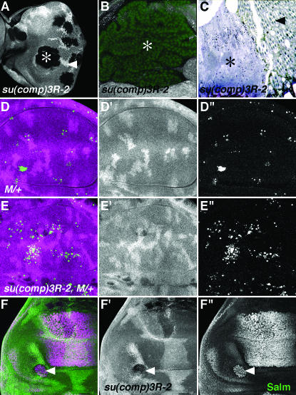
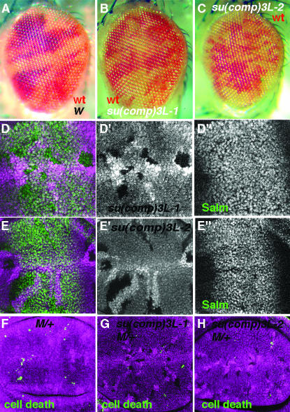
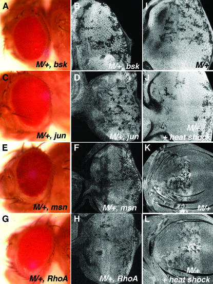
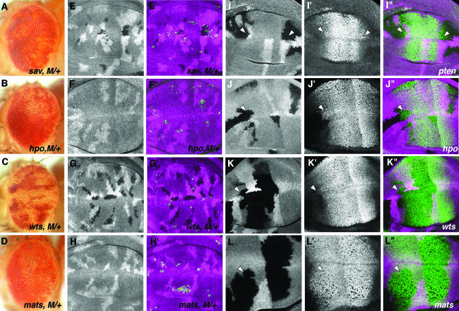
Cell competition is a homeostatic mechanism that regulates the size attained by growing tissues. We performed an unbiased genetic screen for mutations that permit the survival of cells being competed due to haplo-insufficiency for RpL36. Mutations that protect RpL36 heterozygous clones include the tumor suppressors expanded, hippo, salvador, mats, and warts, which are members of the Warts pathway, the tumor suppressor fat, and a novel tumor-suppressor mutation. Other hyperplastic or neoplastic mutations did not rescue RpL36 heterozygous clones. Most mutations that rescue cell competition elevated Dpp-signaling activity, and the Dsmurf mutation that elevates Dpp signaling was also hyperplastic and rescued. Two nonlethal, nonhyperplastic mutations prevent the apoptosis of Minute heterozygous cells and suggest an apoptosis pathway for cell competition . In addition to rescuing RpL36 heterozygous cells, mutations in Warts pathway genes were supercompetitors that could eliminate wild-type cells nearby. The findings show that differences in Warts pathway activity can lead to competition and implicate the Warts pathway, certain other tumor suppressors, and novel cell death components in cell competition, in addition to the Dpp pathway implicated by previous studies. We suggest that cell competition might occur during tumor development in mammals.


Figures

F<sc>igure</sc> 1.—
Figure 1.—
A screen for survival of M/+ cells in a wild-type background. (A) Crossing scheme for screening chromosome arm 2L for mutations; analogous schemes were used to screen the other autosome arms. Males with isogenized FRT40 chromosomes were mutagenized with EMS. The mutagenized chromosomes of interest are depicted in red. Mutagenized males were mated to females carrying both a deficiency that deletes the M(1)Bld gene encoding RpL36 and an FRT40 chromosome bearing a genomic DNA rescue construct (P[RpL36+ w+]). F1 females of the appropriate genotype were screened for surviving white clones. (B) A mosaic eye from the genotype yweyf/+; FRT40/P[RpL36+ w+] FRT40. The adult eye is divided into two genotypes, distinguishable by the presence (P[RpL36+ w+] FRT40/P[RpL36+ w+] FRT40) or absence (FRT40/FRT40) of red pigment. (C) Cell competition eliminated the white M/+ eye cells of the genotype yweyf/Df(1)R194; FRT40/FRT40 from this yweyf/Df(1)R194; FRT40/P[RpL36+ w+] FRT40 fly. (D) A mosaic eye imaginal disc from the genotype yweyf/Df(1)R194; P[RpL36+ w+] FRT40/P[RpL36+ w+] arm-lacZ FRT40, labeled for β-galactosidase expression. The disc contains P[RpL36+ w+]FRT40 homozygous clones that lack β-galactosidase and P[RpL36+ w+] arm-lacZ FRT40 homozygous clones that express it. All cells have the same RpL36 gene dose, so no cell competition occurs. (E) Cell competition eliminated most P[RpL36+ w+] arm-lacZ FRT40 homozygous cells from this yweyf/Df(1)R194; FRT40/P[RpL36+ w+] arm-lacZ FRT40 eye disc, because they lack the P[RpL36+ w+] transgene and are therefore M/+. Some small M/+ clones persist in the posterior part of the eye field, but M/+ cells were almost entirely eliminated from the more anterior parts of the disc. (Anterior is to the left, posterior to the right.) (F) In an adult eye of genotype yweyf/Df(1)R194 ; FRT80/P[RpL36+ w+] FRT80, white cells of the FRT80/FRT80 genotype are eliminated by cell competition (the paler cells visible are the unrecombined heterozygous genotype).(G) In an adult eye of genotype yweyf/Df(1)R194; FRT80 M(3R)/P[RpL36+ w+] FRT80, an unidentified M mutation on the mutagenized chromosome arm 3R renders all cells M/+, irrespective of their dosage of M(1)Bld, so that there is no competition between the 3L recombinant genotypes.
F<sc>igure</sc> 2.—
Figure 2.—
Mutations recovered in a screen for rescue of cell competition. (A–F) These heads contain white eye tissue composed of cells that are RpL36/+ rescued by homozygosity for the following mutations: (A) exNY1, (B) ftNY1, (C) ftNY2, (D) su(comp)3R-1, (E) su(comp)3L-2, and (F) su(comp)3L-1. In the case of su(comp)3R-1 (D), the mutant cells fail to differentiate as eye tissue, resulting in a rough, scarred appearance.
F<sc>igure</sc> 3.—
Figure 3.—
Dpp signaling and apoptosis of M/+ cells in expanded mutants. (A and B) Wing discs containing exNY1 mutant clones induced in the genotype hsflp/+; arm-lacZ FRT40/exNY1 FRT40. Clones that lack β-galactosidase are highlighted by magenta. (A) Salm levels (green) are increased in exNY1 tissue (arrow). This effect is most notable in the posterior part of the Salm expression domain, which corresponds to the region between the presumptive L4 and L5 veins (de Celis and Barrio 2000). This is the region of the wing that is most strongly affected by viable mutations in ex (Boedigheimer and Laughon 1993). In addition, Salm levels are decreased in +/+ tissue (highest levels of β-galactosidase signal are indicated by the arrowhead), compared to heterozygous exNY1/+ tissue. (B) Brk expression (green) is decreased in exNY1 mutant clones (arrow). (C and D) Wing discs containing M/+ clones fixed 48 hr (C) or 72 hr (D) after clone induction (genotype hsflp/M(1)Bld; P[RpL36+ w+] armlacz FRT40/FRT40). M/+ cells lack β-galactosidase (a single representative confocal section is shown in magenta). Apoptotic cells within the disc epithelium are projected in green. There are often more apoptotic cells beneath the disc epithelium, presumably having been expelled. These have not been illustrated or quantified here, as their origin cannot be precisely determined. (C) The majority (≥60%) of CM1-positive cells that could be scored were M/+. Inset shows an enlargement of a single confocal through a typical M/+ clone, so that the genotype of dying cells can be determined more precisely. Two of the three apoptotic cells clearly lack β-galactosidase and so are M/+ (arrows in inset). Both are single cells, surrounded by β-galactosidase-positive neighbors. The third dying cell (arrowhead in inset) may be β-galactosidase positive, although it is difficult to rule out the presence of a M/+ cell. (D) M/+ clones are almost entirely eliminated 72 hr after induction of clones. A few dying cells are left. (E and F) ex, M/+ clones fixed 48 hr (E) or 72 hr (F) after clone induction (genotype hsflp/M(1)Bld; P[RpL36+ w+] armlacz FRT40/exNY1 FRT40). ex, M/+ cells lack β-galactosidase (magenta). Apoptotic cells are labeled in green. (E) The number of apoptotic ex, M/+ cells is similar to the control (compare with C). The ratio of homozygous ex, apoptotic M/+ cells to clone size after 48 hr was compared to that for the M/+ control and was not significantly different. (F) ex, M/+ clones still survive 72 hr after clone induction (contrast with D). The high level of apoptosis indicates that loss of ex accelerates the growth of M/+ cells without protecting them from apoptosis.
F<sc>igure</sc> 4.—
Figure 4.—
Dpp signaling and apoptosis of M/+ cells in fat mutants. (A) ftNY1 mutant clones induced in the genotype hsflp/+; arm-lacZ FRT40/ ftNY1 FRT40. Mutant clones are marked by the absence of β-galactosidase (magenta). Salm levels (green) are increased in ftNY1 clones (arrow). In addition, Salm levels are decreased in +/+ tissue (highest levels of β-galactosidase signal are indicated by an arrowhead).(B) M/+ clones fixed 48 hr after induction (genotype hsflp/M(1)Bld; P[RpL36+ w+] arm-lacZ FRT40/FRT40). M/+ cells that lack β-galactosidase immunofluorescence are in magenta. Only small patches of M/+ cells remain (smaller than their associated twin spots) and apoptotic cells can be seen within these clones (green). (C) ftNY1, M/+ mutant clones fixed 48 hr after induction (genotype hsflp/M(1)Bld; P[RpL36+ w+] arm-lacZ FRT40/ftNY1 FRT40). Clones are smaller than their twin spots and contain dying cells (green). The ratio of homozygous ft, apoptotic M/+ cells to clone size after 48 hr was compared to that for the M/+ control and was not significantly different. (D) ft, M/+ mutant clones fixed 72 hr after clone induction. Few ftNY1, M/+ cells remain.(E–G) Eye imaginal discs were labeled for galactosidase 72 hr after clone induction. In E–G, anterior is to the left and the arrowhead marks the position of the morphogenetic furrow. (E) M/+ clones are almost entirely eliminated; some small clones persist at the posterior edge of the disc. Reciprocal +/+ clones (arrow) show that recombination occurred and M/+ clones must have been eliminated anteriorly to the morphogenetic furrow. (F) exNY1, M/+ clones grow much larger than control clones and are able to survive in much more anterior positions in the eye disc. (G) ftNY1, M/+ clones grow larger and persist more anteriorly than control clones (compare with E).
F<sc>igure</sc> 5.—
Figure 5.—
Dpp signaling, apoptosis of M/+ cells, and differentiation in su(comp)3R-1 mutants. (A) su(comp)3R-1 clones in eye imaginal discs 72 hr after clone induction (gentoype hsflp/M(1)Bld; FRT82/FRT82 P[RpL36+ w+] arm-lacZ). Clones (asterisk) are larger than twin spots (arrowhead).(B) su(comp)3R-1 mutant clone (asterisk) in the wing; mutant tissue folds to accommodate the dramatic overgrowth. Nuclei of all cells are labeled in green with DRAQ-5. (C) In a section through the adult eye containing su(comp)3R-1 mutant (asterisk) and wild-type (arrowhead) tissue, the mutant tissue is amorphous and lacks the regular array of rhabdomeres. (D) Control M/+ clones in wing discs labeled 48 hr after clone induction (gentoype hsflp/M(1)Bld; FRT82/FRT82 P[RpL36+ w+] arm-lacZ). M/+ cells lack β-galactosidase (shown in magenta), but few are seen. Apoptotic cells are in green. (E) su(comp)3R-1, M/+ clones in wing discs labeled 48 hr after clone induction (genotype hsflp/M(1)Bld; FRT82 3R-12/FRT82 P[RpL36+ w+] arm-lacZ). 3R-12, M/+ clones are present, despite abundant cell death. (F) Salm expression (green and F′′) is upregulated in su(comp)3R-1 clones (arrowhead) (genotype hsflp/+; FRT82 su(comp)3R-1 /FRT82 arm-lacZ).
F<sc>igure</sc> 6.—
Figure 6.—
Dpp signaling and apoptosis of M/+ cells in su(comp)3L-1 and su(comp)3L-2 mutants. (A) The eye is divided roughly equally into red and white tissue in a control (genotype y w Ey-FLP; FRT80/w+ FRT80). (B and C) Proportions of mutant (white) and control (red) tissue are also similar when clones are homozygous for su(comp)3L-1 (B) or su(comp)3L-2(C). (D) Salm levels in the wing disc (D″ and green in D) are not affected in su(comp)3L-1 clones. (E) Salm levels in the wing disc (D″ and green in D) are are not affected in su(comp)3L-2 clones. (F) M/+ clones (those lacking the magenta labeling for β-galactosidase) 48 hr after induction (genotype hsflp/M(1)Bld; P[RpL36+ w+] arm-lacZ FRT80/FRT80). Most have been lost; only a few apoptotic corpses remain (green). (G) M/+, su(comp)3L-1 clones persist with little apoptosis. (H) M/+, su(comp)3L-1 clones persist with little apoptosis.
F<sc>igure</sc> 7.—
Figure 7.—
Dpp signaling and apoptosis of M/+ cells in Dsmurf mutants. (A) No white M/+ cells remain in the y w Ey-FLP/Df(1)R194; FRT42/FRT42 P[RpL36+ w+] P[armLacZ w+] eye. (B) DsmurfKG07014, M/+ cells do survive in the adult eye (genotype y w Ey-FLP/Df(1)R194; FRT42 DsmurfKG07014/FRT42 P[RpL36+ w+]). Because the DsmurfKG07014 mutation is caused by a w+ P-element insertion, DsmurfKG07014 homozygous cells are the more pigmented genotype (e.g., arrowhead).(C) Pigmented DsmurfKG07014 homozygous clones (e.g., arrowhead) predominate in a mosaic with wild-type cells (genotype y w Ey-FLP/+; FRT42 DsmurfKG07014/FRT42).(D) Salm protein levels (green) are upregulated in DsmurfKG07014 homozygous cells (those lacking β-galactosidase are in magenta). Both subtle increases in expression level and lateral expansion of the expression domain occur (e.g., arrows). DsmurfKG07014 hyperplasia is less certain in the wing than in the eye, perhaps reflecting differing growth effects of Dpp signaling in distinct regions of the wing disc (Rogulja and Irvine 2005). (E and F) M/+ clones are small and apoptotic after 48 hr (E) and mostly eliminated after 72 hr (F) (genotype hsflp/M(1)Bld; FRT42 P[RpL36+ w+] arm-lacZ /FRT42). (G and H). M/+, DsmurfKG07014 clones are small and apoptotic 48 hr after induction (G), but many survive 72 hr after induction (H) (genotype hsflp/M(1)Bld; FRT42 P[RpL36+ w+] arm-lacZ /FRT42 DsmurfKG07014). (I) exNY1 Mad10, M/+ clones can survive in the adult eye (e.g., arrows) (genotype y w Ey-FLP/Df(1)R194; P[RpL36+ w+] P[armLacZ w+] FRT40/exNY1 Mad10 FRT40). Some clones also have differentiation defects. Control M/+ clones are shown in Figure 1C.
F<sc>igure</sc> 8.—
Figure 8.—
The JNK pathway and heat shock (A–H). M/+ clones also homozygous for JNK components were induced using eyFLP. None of the JNK mutations protected M/+ clones in adults (A: bsk2; C: jun2; E: msn102; and G: RhoABH; compare the negative control in Figure 1C and the positive controls in Figure 2). None rescued in eye discs either (B: bsk2; D: jun2; F: msn102; and H: RhoABH; compare the negative control in Figure 1E and the positive controls in Figure 4, F and G). None of the mutations affected clonal growth adversely by themselves (data not shown). (I–L) M/+ clones were induced continuously in eyes by eyFLP (I and J) or in anterior wings by dppGal4>UAS:FLP (K and L). M/+ clones were eliminated more efficiently in tissues that had been heat-shocked 72 hr earlier (J and L) than in those that had not (I and K).
F<sc>igure</sc> 9.—
Figure 9.—
A subset of hyperplastic tumor suppressors prevent cell competition or affect Dpp signaling. (A–D) Mutations in the Warts pathway allow survival of M/+ cells. The following mutations allowed white, M/+ clones to survive in the adult eye: (A) sav3, (B) hpoMGH4, (C) wtsMGH1, and (D) matsroo. (E–H) M/+ clones also homozygous for sav3 (E), hpoMGH4 (F), wtsMGH1 (G), and matsroo (H) were fixed 48 hr after clone induction. Apoptosis (green) is reduced by sav and wts compared to controls, but not by hpo or mats. See Figure 5 for FRT82 control for wts, mats, and sav. The ratio of homozygous mutant, apoptotic cells to clone size was compared, except that too few matsroo, M//+ cells or hpoMGH4, M/+ cells were found for quantification. (I–L) Salm protein levels are upregulated in mutant cells: PTENdj189 (I), hpoMGH4 (J), wtsMGH1 (K), and matsroo(L). Note either an increase in the intensity of labeling relative to the adjacent tissue or the lateral expansion of label within the clone (arrowheads). Clones of mutant cells are marked by the absence of the β-galactosidase label (magenta, I′′–L′′). Salm protein is labeled green in merges (I′′–L′′). Homozygous mats cells have notably larger nuclei (L′), but this cannot account for the intensity of Salm labeling, which is increased in single confocal sections.
F<sc>igure</sc> 10.—
Figure 10.—
Warts pathway mutants compete with wild type. All panels show cell death (green) in discs containing clones of homozygous mutant cells lacking β-galactosidase (magenta). Some other cells die next to clones for each of the Warts pathway mutants (arrowheads). In addition, for ex, some wild-type cells (+/+, twice the β-galactosidase level) die next to heterozygous ex/+ cells (example in A).

Similar articles

Cited by

References

    1. Adler, P. N., J. Charlton, K. H. Jones and J. Liu, 1994. The cold-sensitive period for frizzled in the development of wing hair polarity ends prior to the start of hair morphogenesis. Mech. Dev. 46: 101–107. - PubMed
    1. Adler, P. N., J. Charlton and J. Liu, 1998. Mutations in the cadherin superfamily member gene dachsous cause a tissue polarity phenotype by altering frizzled signaling. Development 125: 959–968. - PubMed
    1. Berger, J., T. Suzuki, K. A. Senti, J. Stubbs, G. Schaffner et al., 2001. Genetic mapping with SNP markers in Drosophila. Nat. Genet. 29: 475–481. - PubMed
    1. Bilder, D., and N. Perrimon, 2000. Localization of apical epithelial determinants by the basolateral PDZ protein Scribble. Nature 403: 676–680. - PubMed
    1. Blaumueller, C. M., and M. Mlodzik, 2000. The Drosophila tumor suppressor expanded regulates growth, apoptosis, and patterning during development. Mech. Dev. 92: 251–262. - PubMed

Publication types

Substances