

Hippocampal infolding angle changes during brain development assessed by prenatal MR imaging
- PMID: 17110674
- PMCID: PMC7977211
Hippocampal infolding angle changes during brain development assessed by prenatal MR imaging
Abstract
Background and purpose: Epileptic syndromes or neurodevelopmental delay may be associated with congenital anomalies of the shape or the orientation of the hippocampus. Scarce data are available about quantitative hippocampal developmental changes during fetal life, in particular about the progressive rotational changes of the hippocampal infolding angle (HIA), which can be considered a hallmark of hippocampal development. We hypothesized that prenatal MR imaging could demonstrate the progressive rotation of the hippocampus, providing quantitative data by means of the HIA determination.
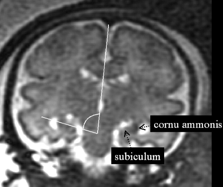
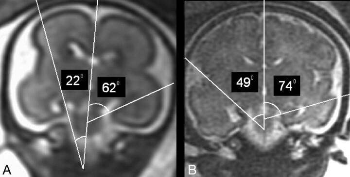
Methods: We retrospectively selected 62 fetal MR imaging cases with normal brain at prenatal and postnatal imaging. The gestational age ranged from 20 to 37 weeks. The coronal section encompassing the pons was used to perform the measurement of HIA. HIA was defined as the angle between the line connecting the lateral margin of the cornu ammonis with the medial superior margin of the subiculum and the line passing through the midline structures.
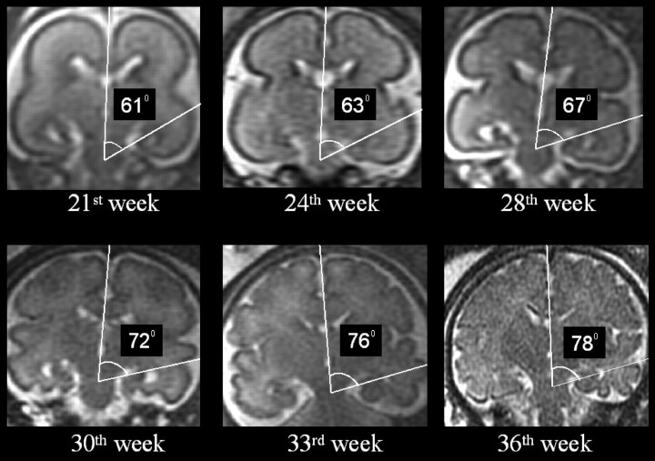
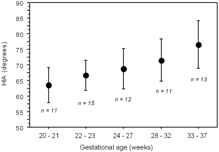
Results: A significant positive correlation was found between the HIA value and the gestational age. The HIA was generally below 70 degrees before the gestational week 25 and above 70 degrees after week 30.
Conclusion: Prenatal MR imaging allowed the progressive rotation of hippocampus to be detected during fetal life, providing normative data about HIA changes. These data could support further investigations to assess how fetal HIA anomalies might affect postnatal neurologic outcome.
Figures

References
-
- Baulac M, De Grissac N, Hasboun D, et al. Hippocampal developmental changes in patients with partial epilepsy: magnetic resonance imaging and clinical aspects. Ann Neurol 1998;44:223–33 - PubMed
-
- Okada Y, Toshinori K, Iwai K, et al. Evaluation of hippocampal infolding using magnetic resonance imaging. NeuroReport 2003;14:1405–09 - PubMed
-
- Bernasconi N, Kinay D, Andermann F, et al. Analysis of shape and positioning of the hippocampal formation: an MRI study in patients with partial epilepsy and healthy controls. Brain 2005;128:2442–52 - PubMed
MeSH terms
LinkOut - more resources
Full Text Sources
Medical