

Hyperintensity of the middle cerebellar peduncles on fluid-attenuated inversion recovery imaging: variation with age and implications for the diagnosis of multiple system atrophy
- PMID: 17110685
- PMCID: PMC7977213
Hyperintensity of the middle cerebellar peduncles on fluid-attenuated inversion recovery imaging: variation with age and implications for the diagnosis of multiple system atrophy
Abstract
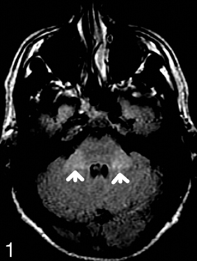
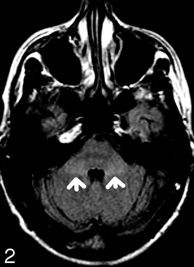
Background and purpose: T2 hyperintensity of the middle cerebellar peduncle (MCP) is described in a number of diseases, including multiple system atrophy (MSA). We hypothesize that mild MCP hyperintensity on fluid-attenuated inversion recovery (FLAIR) imaging can be a normal finding. To our knowledge, a detailed study of the prevalence of this finding in various age groups with the FLAIR sequence has not been described.
Methods: One hundred twenty-two patients underwent an axial FLAIR examination of the brain as part of either a hearing loss or tinnitus work-up (ie, to exclude an acoustic neuroma or a retrocochlear cause). Subjects aged 15-78 years were included to reflect an even spread through the decades and were divided into 6 age groups. A radiologist and an MR imaging fellow graded the examinations subjectively, blinded to age: 0 for normal or 1 for the presence of MCP hyperintensity if the increased signal intensity was greater than that of adjacent pons and cerebellar white matter. Spearman rank correlation test of MCP hyperintensity with age and analysis of variance (ANOVA) were performed.
Results: Of 122 patients, we identified 17 with MCP FLAIR hyperintensity. None of these patients had a clinical condition that could cause MCP hyperintensity. MCP hyperintensity did not show a statistically significant correlation with age (r = 0.05, P = .62). Patients were divided into 6 age groups, and ANOVA showed no statistically significant difference in the incidence of MCP hyperintensity between different age groups (P = .95). However, results were highly reproducible with excellent interobserver correlation (r = 0.97, P < .001).
Conclusions: Mild MCP FLAIR hyperintensity can occur normally, and this finding shows no relationship with age.
Figures
References
-
- Seppi K, Schocke MF, Wenning GK, et al. How to diagnose MSA early: the role of magnetic resonance imaging. J Neural Transm 2005;112:1625–34 - PubMed
-
- Schrag A, Good CD, Miszkiel K, et al. Differentiation of atypical parkinsonian syndromes with routine MRI. Neurology 2000;55:1239–40 - PubMed
-
- Savoiardo M. Differential diagnosis of Parkinson’s disease and atypical parkinsonian disorders by magnetic resonance imaging. Neurol Sci 2003;24:S35–S37 - PubMed
-
- Lee EA, Cho HI, Kim SS, et al. Comparison of magnetic resonance imaging in subtypes of multiple system atrophy. Parkinsonism Relat Disord 2004;10:363–68 - PubMed
MeSH terms
LinkOut - more resources
Full Text Sources
Medical
Miscellaneous