

In vivo detection of cortical plaques by MR imaging in patients with multiple sclerosis
- PMID: 17110688
- PMCID: PMC7977209
In vivo detection of cortical plaques by MR imaging in patients with multiple sclerosis
Abstract
Background and purpose: In vivo detection of cortical lesions in patients with multiple sclerosis (MS) by MR imaging is hampered by several factors. Among them is the low contrast between small cortical lesions and surrounding cortical gray matter offered by present techniques.
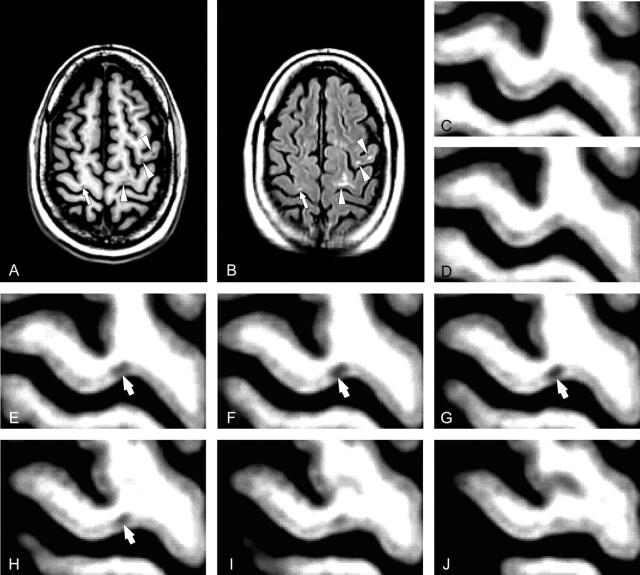
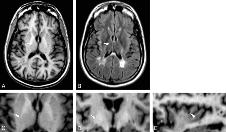
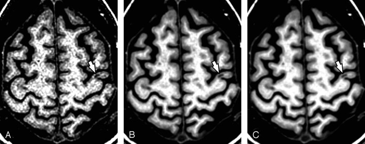
Methods: T1-weighted 3D spoiled gradient-recalled-echo (SPGR) volumes and 2D fluid-attenuated inversion recovery (FLAIR) sequences of 22 patients with MS who had 12 monthly brain MR imaging examinations at 1.5T, using a quadrature head coil, were retrospectively analyzed. These serial studies were coregistered and averaged to generate a single high signal-to-noise ratio (SNR) mean image, which was used to identify cortical lesions. The means of 12 FLAIRs and SPGRs from 14 age- and sex-matched healthy volunteers were analyzed as well.
Results: No cortical lesions were found on images of healthy subjects. Eighty-six cortical lesions were identified in 13 (59.1%) patients, predominantly in the frontal lobe (73.3%); 23.3% of cortical lesions lay entirely in the cortex, whereas the remaining lesions invaded the white matter underneath.
Conclusion: Averaging multiple SPGRs created a single high SNR volume, allowing identification of cortical lesions. Because data were obtained monthly for 1 year, the average image does not account for transient lesion activity. However, for cortical lesions that remained stable during this time, the findings are valid in demonstrating the importance of high SNR images for detecting cortical brain abnormalities in MS.
Figures
References
-
- Kidd D, Barkhof F, McConnell R, et al. Cortical lesions in multiple sclerosis. Brain 1999;122:17–26 - PubMed
-
- Peterson JW, Bo L, Mork S, et al. Transected neuritis, apoptotic neurons, and reduced inflammation in cortical multiple sclerosis lesions. Ann Neurol 2001;50:389–400 - PubMed
-
- Bo L, Vedeler CA, Nyland HI, et al. Subpial demyelination in the cerebral cortex of multiple sclerosis patients. J Neuropathol Exp Neurol 2003;62:723–32 - PubMed
-
- Brink BP, Veerhuis R, Breij EC, et al. The pathology of multiple sclerosis is location-dependent: no significant complement activation is detected in purely cortical lesions. J Neuropathol Exp Neurol 2005;64:147–55 - PubMed
Publication types
MeSH terms
Grants and funding
LinkOut - more resources
Full Text Sources
Medical