

Case Reports
MR features of intraocular ectopic lacrimal tissue
Affiliations
- PMID: 17110692
- PMCID: PMC7977227
Item in Clipboard
Case Reports
MR features of intraocular ectopic lacrimal tissue
AJNR Am J Neuroradiol.
2006 Nov-Dec.
Abstract
A 2-year-old girl who had a 3-day history of swelling in her right eye presented with a case of intraocular ectopic lacrimal tissue. MR imaging findings and possible causes of the ectopic lacrimal tissue in the globe, including embryology, are reviewed. The differential diagnoses of other intraocular masses and the imaging features that can be of help in making a diagnosis are also discussed.
Figures
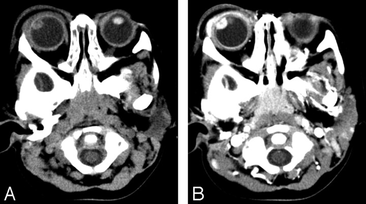
A, Precontrast axial orbital CT revealed a soft tissue attenuation ovoid mass in the superolateral portion of right eye. B, Postcontrast axial orbital CT scan showed intense enhancement of an ovoid mass. Swelling in soft tissue around right preseptal area and ipsilateral lacrimal gland suggested orbital cellulitis with lacrimal adenitis.

T1- and T2-weighted MR images revealed a circumscribed ringlike mass in superolateral aspect of right eye. The central portion of the mass was low signal intensity relative to vitreous on coronal T1WI (A) and high signal intensity on T2WI (B). The peripheral portion of mass revealed slightly high signal intensity on T1WI and low signal intensity on T2WI. Coronal gadolinium-enhanced T1WI (C) showed strong and homogenous enhancement in peripheral portion of mass in continuity with the enhancing uveal tract.
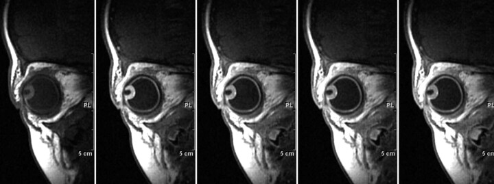
Dynamic enhanced T1-weighted MR sagittal scans showed early and homogenous strong enhancement in the mass.
References
-
- Green WR, Zimmerman LE. Ectopic lacrimal tissue tissue. Report of eight cases with orbital involvement. Arch Ophthalmol 1967;78:318–27 - PubMed
-
- Puech PM. Adénome de la choroïde. J Med Bord Sud-Ouest 1887;31:342
-
- Rush A, Leone CR Jr. Ectopic lacrimal gland cyst of the orbit. Am J Ophthalmol 1981;92:198–201 - PubMed
-
- Bruce GM. Aberrant glandular tissue in the iris. Trans Am Acad Ophthalmol Otolaryngol 1952;56:47–51 - PubMed
-
- Christensen L, Anderson ED. Aberrant intraocular adenomata and epithelization of the anterior chamber. AMA Arch Ophthalmol 1952;48:19–29 - PubMed
Publication types
MeSH terms
LinkOut - more resources
Full Text Sources
Medical