. 2006 Nov-Dec;27(10):2204-9.

Imaging and clinical characteristics of temporal bone meningioma

Affiliations

Imaging and clinical characteristics of temporal bone meningioma

B E Hamilton et al. AJNR Am J Neuroradiol. 2006 Nov-Dec.

Abstract

Background and purpose: Imaging characteristics of temporal bone meningioma have not been previously reported in the literature. CT and MR imaging findings in 13 cases of temporal bone meningioma are reviewed to define specific imaging features.

Methods: A retrospective review of our institutional case archive revealed 13 cases of histologically confirmed temporal bone meningioma. CT and MR imaging studies were reviewed to characterize mass location, vector of spread, bone changes, enhancement characteristics, and intracranial patterns of involvement. Clinical presenting signs and symptoms were correlated with imaging findings.

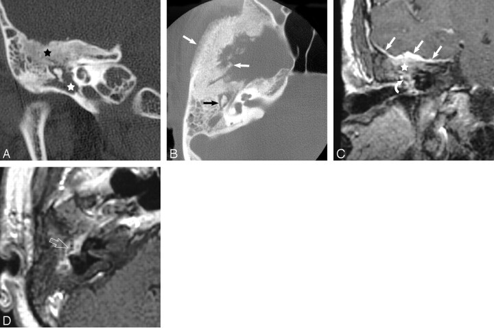
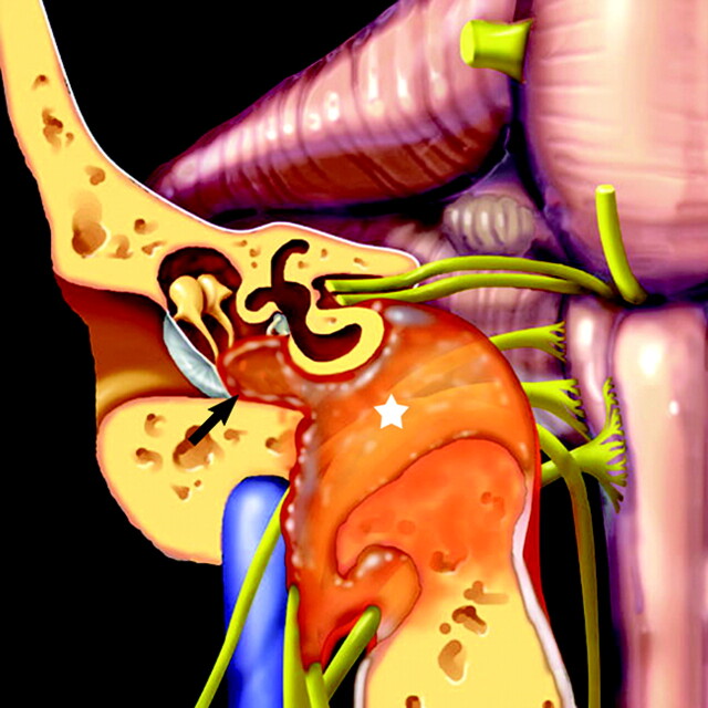
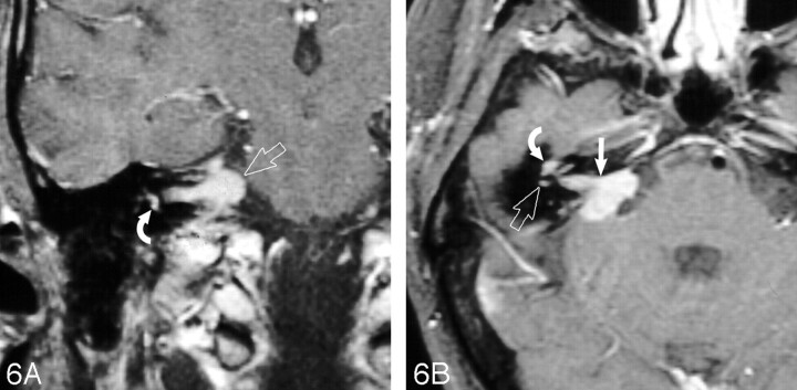
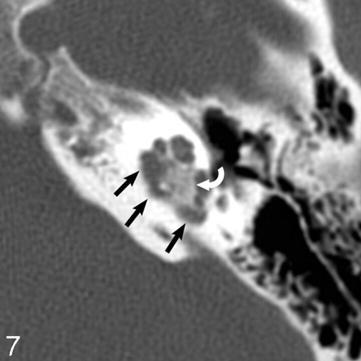
Results: Thirteen temporal bone meningiomas were reviewed in 8 women and 5 men, aged 18-65 years. Meningiomas were stratified into 3 groups on the basis of location and tumor vector of spread. There were 6 tegmen tympani, 5 jugular foramen (JF), and 2 internal auditory canal (IAC) meningiomas. Tegmen tympani and JF meningiomas were characterized by spread to the middle ear cavity. IAC meningiomas, by contrast, spread to the cochlea and vestibule. Hearing loss was the most common clinical presenting feature in all cases of temporal bone meningioma (10/13). The presence of tumor adjacent to the ossicles strongly correlated with conductive hearing loss (7/9).

Conclusion: Meningioma involving the temporal bone is rare. Three subgroups of meningioma exist in this location: tegmen tympani, JF, and IAC meningioma. Tegmen tympani and JF meningiomas spread to the middle ear cavity. IAC meningiomas spread to intralabyrinthine structures. Conductive hearing loss is commonly seen in these patients and can be surgically correctable.


Figures

Fig 1.
Fig 1.
Coronal graphic depicts characteristic findings in tegmen tympani meningioma. Tumor arises in the middle cranial fossa and spreads inferomedially through the tegmen tympani to the middle ear cavity. En plaque intracranial enhancement is typical (straight white arrows). Ossicles are encased by tumor (curved white arrow), without destruction (reprinted with permission from Amirsys).
Fig 2.
Fig 2.
A, Coronal unenhanced CT scan reveals typical thickening and trabecular hyperostosis of the tegmen tympani (black star). Note the soft-tissue mass in the middle ear cavity (white star), which encases the ossicles but lacks erosive or destructive change. B, Axial unenhanced CT scan shows ossicular encasement without erosive or destructive changes (black arrow). The involved calvaria (white arrows) is characterized by preserved internal architecture (trabecular hyperostosis) and irregularity of the inner table. C, Coronal contrast-enhanced MR image shows typical en plaque enhancement along the floor of the middle cranial fossa (straight white arrows) in a tegmen tympani meningioma. Note thickened enhancing tegmen (white star) and enhancing tissue surrounding the ossicular chain (curved white arrow). D, Axial contrast-enhanced MR image of the tegmen tympani meningioma shows tumor enhancement in the middle ear cavity. Note the intact appearance of the ossicular chain (open white arrow), seen through the enhancement.
Fig 3.
Fig 3.
Coronal graphic depicts findings in JF meningioma: location primary to the JF (white star) with superolateral vector of spread of the soft-tissue mass into the middle ear cavity (black arrow) (reprinted with permission from Amirsys).
Fig 4.
Fig 4.
A, Coronal unenhanced CT scan shows a mixed permeative/sclerotic appearance of the involved skull base (white star) that is typical for JF meningioma. Bony changes mildly narrow the mastoid segment of the facial nerve canal (open white arrow). B, Axial unenhanced CT scan again demonstrates the characteristic mixed permeative/sclerotic appearance and centrifugal involvement of the skull base (black star). Note intact-appearing cortical margins (black arrows). Although not a common finding, calcification of the soft-tissue mass (white arrow) is a fairly specific finding of meningioma when present. C, Coronal enhanced MR image demonstrates an enhancing mass primarily located in the jugular foramen (white arrow), with centrifugal involvement of the surrounding skull base and superolateral soft-tissue spread to the middle ear cavity (black star). Note intense intraosseous enhancement relative to the extracranial component and largely preserved intrinsic bony architecture (black arrows). D, Axial enhanced MR image demonstrates an intensely enhancing mass surrounding the intact-appearing ossicular chain (open white arrow). Note the intracranial enhancing dural tail (straight white arrow). Secondary extension to the internal auditory canal (curved white arrow) is also present in this case.
Fig 5.
Fig 5.
Coronal graphic depicts findings in IAC meningioma: tumor in the cochlea and vestibular apparatus (black arrows). Note the intracranial component in the IAC and cerebellopontine angle (black star) (reprinted with permission from Amirsys).
Fig 6.
Fig 6.
A, Coronal enhanced MR image of an IAC meningioma demonstrates intense enhancement of the intralabyrinthine structures (curved white arrow). The mass extends from the IAC and cerebellopontine angle (open white arrow). B, Axial enhanced MR image of an IAC meningioma demonstrates intense enhancement of the cochlea (curved white arrow) and vestibular apparatus (open white arrow). The large IAC and cerebellopontine angle enhancing mass (straight white arrow) are clues to the diagnosis.
Fig 7.
Fig 7.
Axial unenhanced CT scan demonstrates pericochlear bone lucency (straight black arrows). Calcification of the mass (curved white matter) suggests the diagnosis of meningioma, as seen in other locations.

References

    1. Prayson RA. Middle ear meningiomas. Ann Diag Pathol 2000;4:149–53 - PubMed
    1. Macdonald AJ, Salzman KL, Harnsberger HR, et al. Primary jugular foramen meningioma: imaging appearance and differentiating features. AJR Am J Roentgenol 2004;182:373–77 - PubMed
    1. Thompson LD, Bouffard JP, Sandberg GD, et al. Primary ear and temporal bone meningiomas: a clinicopathologic study of 36 cases with a review of the literature. Mod Pathol 2003;16:236–45 - PubMed
    1. Aho TR, Daspit DP, Dean BL, et al. Intralabyrinthine meningioma. AJNR Am J Neuroradiol 2003;24:1642–45 - PMC - PubMed
    1. Rosahl SK, Mirzayan MJ, Samii M. Osteolytic intra-osseous meningiomas: illustrated review. Acta Neurochir (Wien) 2004;146:1245–49. Epub 2004 Sep 9 - PubMed