. 2006 Nov 29;25(23):5516-26.
doi: 10.1038/sj.emboj.7601432. Epub 2006 Nov 16.

Structural mechanism of RPA loading on DNA during activation of a simple pre-replication complex

Affiliations

Structural mechanism of RPA loading on DNA during activation of a simple pre-replication complex

Xiaohua Jiang et al. EMBO J. .

Abstract

We report that during activation of the simian virus 40 (SV40) pre-replication complex, SV40 T antigen (Tag) helicase actively loads replication protein A (RPA) on emerging single-stranded DNA (ssDNA). This novel loading process requires physical interaction of Tag origin DNA-binding domain (OBD) with the RPA high-affinity ssDNA-binding domains (RPA70AB). Heteronuclear NMR chemical shift mapping revealed that Tag-OBD binds to RPA70AB at a site distal from the ssDNA-binding sites and that RPA70AB, Tag-OBD, and an 8-nucleotide ssDNA form a stable ternary complex. Intact RPA and Tag also interact stably in the presence of an 8-mer, but Tag dissociates from the complex when RPA binds to longer oligonucleotides. Together, our results imply that an allosteric change in RPA quaternary structure completes the loading reaction. A mechanistic model is proposed in which the ternary complex is a key intermediate that directly couples origin DNA unwinding to RPA loading on emerging ssDNA.


Figures

Figure 1
Figure 1
Domain organization of RPA with Tag-binding regions shaded. Rectangles depict OB-folds, of which four are ssDNA-binding domains (DBDs; A–D); an oval indicates the winged-helix–turn–helix of RPA32C (Mer et al, 2000); a circled P depicts the phosphorylated region of RPA32. A triple arrow symbolizes the association of subunits through a three-helix bundle (Bochkareva et al, 2002), and hexagons denote Tag hexamers.
Figure 2
Figure 2
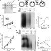
Tag interaction with RPA70AB actively loads human RPA onto ssDNA during initiation of SV40 DNA replication. (A) Purified hRPA, yRPA, and hRPAy32C chimera were visualized by SDS–PAGE and Coomassie stain. PM, protein mass marker. (B, C) Filter binding assays were used to compare the activity of the indicated RPAs in binding to radiolabeled dT30. (D, E) SV40 monopolymerase assays were performed with a suboptimal amount of human RPA (0.2 mg) (lane 1) supplemented with up to five-fold greater amounts of human RPA (lanes 2–4) and either (D) yeast RPA or (E) hRPAy32C (lanes 5–8) as indicated. Reaction products were visualized by denaturing gel electrophoresis and autoradiography (left), and quantified by scintillation counting (right). Negative control reactions lacked Tag, polymerase alpha-primase, or human RPA as indicated (lanes 9–11).
Figure 3
Figure 3
Tag-OBD protects RPA70AB from proteolytic digestion. RPA70AB was incubated with trypsin for the indicated time periods, either alone (A) or in the presence of Tag-OBD (B) d(C)8 (C), or d(C)8 and Tag-OBD (D). Digestion products were visualized by SDS–PAGE and Coomassie staining.
Figure 4
Figure 4
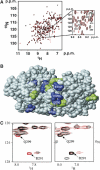
Structural mapping of RPA70AB-binding site for Tag-OBD. (A) Chemical shift mapping of Tag-OBD-binding site on RPA70AB. Overlay of 15N-1H HSQC spectra of 15N-enriched RPA70AB in the absence (black) and presence of Tag-OBD (red). The inset shows some of the residues that are perturbed or broadened upon interaction with Tag-OBD. (B) Molecular surface of RPA70AB with residues whose chemical shifts are perturbed upon binding of Tag-OBD colored in blue (strongest effects) and green (other significant effects). (C) Formation of a stable ternary complex of RPA70AB with d(C)8 and Tag-OBD. The left panel shows a region from the 15N-1H HSQC spectra of the binary complex of 15N-enriched RPA70AB with d(C)8 in the absence (black) and presence of Tag-OBD (red). The right panel shows a region from the 15N-1H HSQC spectra of the binary complex of 15N-enriched RPA70AB with Tag-OBD in the absence (black) and presence of d(C)8 Tag-OBD (red).
Figure 5
Figure 5
Structural mapping of Tag-OBD-binding site on RPA70AB. (A) Chemical shift perturbation analysis of the Tag-OBD-binding site on RPA70AB. Overlay of 15N-1H HSQC spectra of 15N-enriched Tag-OBD in the absence (black) and presence of RPA70AB (red). A expansion of a small region is shown, highlighting some of the residues that are perturbed or broadened upon interaction with RPA70AB. (B, C) Molecular surface diagrams of the electrostatic potential of Tag-OBD and RPA70AB, respectively, with blue for positive charge and red for negative charge. The large yellow circle in (B) highlights the contiguous binding region composed of regions F151-T155, F183-H187, and H203-A207. The three prominent Arg residues providing the bulk of the basic character of this region are labeled. The small yellow circles in (C) highlight the six acidic residues that provide the acidic character to the Tag-OBD-binding site on RPA70AB. The ssDNA in the RPA70AB structure is colored yellow. (D) A charge reversal mutation in Tag-OBD reduces Tag binding to RPA70AB. Wild-type (lanes 1, 3, 4) and R154E mutant Tag (lanes 2, 5, 6) adsorbed to antibody beads were incubated with 5 or 10 μg of RPA70AB as indicated. Proteins bound to the beads were separated by SDS–PAGE and visualized by Western blotting with Mab70C against RPA (top panel) or Pab101 against Tag (lower panel). Antibody beads lacking Tag did not bind RPA70AB (lane 7). Lane 8 shows 200 ng of input RPA70AB. (E) Quantification of bound RPA70AB in lanes 3–6 of (D) after subtraction of background in lane 7.
Figure 6
Figure 6
Transient Tag binding to RPA facilitates ssDNA binding of RPA. (A) Tag bound to Pab101-protein G beads was incubated with RPA (8.6 pmol) or without RPA (con). After washing the beads, increasing amounts of oligonucleotides dT30, dT15, or dT8 (0, 1, 2, 4, 9, 17, or 34 pmol) were added. After 1 h, the amount of RPA that remained bound to Tag was visualized by SDS–PAGE and Western blot with anti-RPA70 antibody. Input: 5% of the RPA added to samples. (B) RPA in the indicated amounts (pmol) was pre-incubated with ∼3 pmol of radiolabeled dT30 for 10 min at 25°C and then, after addition of the indicated amounts of Tag (pmol of hexamer), for another 15 min at 37°C. Protein–DNA complexes were detected by native gel electrophoresis and autoradiography. The migration of Tag-dT30 complex (Supplementary Figure 3) is indicated. W, wells of the gel. (C) RPA (9 pmol) was incubated with ∼3 pmol of dT30, followed by addition of 3.5 pmol of Tag hexamer and, after 5 min, monoclonal antibody (0.5, 2.5, or 5 μg) against Tag (T), influenza hemagglutinin (N), or RPA32C (R). Complexes were visualized as in (B). (D) The indicated amounts (pmol) of yeast RPA (yRPA) were incubated with ∼3 pmol of radiolabeled dT30 in the presence of Tag hexamer (pmol) as indicated. Complexes were analyzed as in (B). (E) Increasing amounts (0.5 or 1 pmol of hexamer) of wild-type (lanes 3 and 4) or mutant R154E Tag (lanes 6 and 7) were added to RPA (2 pmol) that had been pre-incubated with radiolabeled dT30 as indicated, and protein–DNA complexes were analyzed by native gel electrophoresis and autoradiography as in (B).
Figure 7
Figure 7
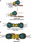
Proposed mechanism for coupling activation of the SV40 pre-replication complex with RPA loading on ssDNA. (A) An active Tag hexamer (OBD and N-terminus in gold; helicase domains in turquoise) translocating 3′ to 5′ on the red strand (dotted line) and displacing the blue strand is proposed to form a ternary complex, in which Tag-OBD binds to RPA70AB (orange) associated with 8–10 nucleotides of ssDNA. As more DNA is unwound, RPA extends into the 30-nucleotide binding mode on ssDNA and releases Tag-OBD. (B) Tag monomers recognize four specific binding sites (black arrows) in the origin DNA, nucleating double hexamer assembly and inducing an ssDNA bubble at the flanking EP sequence and distortion at AT. The depicted path of DNA through the Tag double hexamer is speculative (Gai et al, 2004b; Li et al, 2003; Valle et al, 2006). (C) Remodeling of the Tag N-terminus and Tag-OBD (Meinke et al, 2006; Valle et al, 2006) in the presence of human RPA is proposed to facilitate formation of a ternary complex composed of RPA70AB bound to an ssDNA bubble and to a basic OBD surface exposed on the exterior of each hexamer, analogous to that shown in (A). As the helicase translocates (dotted lines) and more ssDNA emerges, RPA would extend into the 30-nucleotide binding mode on ssDNA (orange arrows) and dissociate from Tag, completing the loading cycle.

References

    1. Alexandrov AI, Botchan MR, Cozzarelli NR (2002) Characterization of Simian Virus 40 T-antigen double hexamers bound to a replication fork. The active form of helicase. J Biol Chem 277: 44886–44897 - PubMed
    1. Arunkumar AI, Klimovich V, Jiang X, Ott RD, Mizoue L, Fanning E, Chazin WJ (2005) Insights into hRPA32 C-terminal domain-mediated assembly of the simian virus 40 replisome. Nat Struct Mol Biol 12: 332–339 - PMC - PubMed
    1. Arunkumar AI, Stauffer ME, Bochkareva E, Bochkarev A, Chazin WJ (2003) Independent and coordinated functions of replication protein A tandem high affinity single-stranded DNA binding domains. J Biol Chem 278: 41077–41082 - PubMed
    1. Bastin-Shanower SA, Brill SJ (2001) Functional analysis of the four DNA binding domains of replication protein A. The role of RPA2 in ssDNA binding. J Biol Chem 276: 36446–36453 - PMC - PubMed
    1. Bhattacharya S, Arunkumar AI, Sullivan SL, Botuyan M-V, Arrowsmith CH, Chazin WJ (2004) Letter to the editor: 1H, 13C and 15N assignments of single-stranded DNA binding domains from the 70 kDa subunit of human replication protein A. J Biomol NMR 28: 195–196 - PubMed

Publication types

Substances