. 2007 Jan;35(1):1-18.
doi: 10.1007/s10439-006-9212-7. Epub 2006 Nov 8.

Coupling of a 3D finite element model of cardiac ventricular mechanics to lumped systems models of the systemic and pulmonic circulation

Affiliations

Coupling of a 3D finite element model of cardiac ventricular mechanics to lumped systems models of the systemic and pulmonic circulation

Roy C P Kerckhoffs et al. Ann Biomed Eng. 2007 Jan.

Abstract

In this study we present a novel, robust method to couple finite element (FE) models of cardiac mechanics to systems models of the circulation (CIRC), independent of cardiac phase. For each time step through a cardiac cycle, left and right ventricular pressures were calculated using ventricular compliances from the FE and CIRC models. These pressures served as boundary conditions in the FE and CIRC models. In succeeding steps, pressures were updated to minimize cavity volume error (FE minus CIRC volume) using Newton iterations. Coupling was achieved when a predefined criterion for the volume error was satisfied. Initial conditions for the multi-scale model were obtained by replacing the FE model with a varying elastance model, which takes into account direct ventricular interactions. Applying the coupling, a novel multi-scale model of the canine cardiovascular system was developed. Global hemodynamics and regional mechanics were calculated for multiple beats in two separate simulations with a left ventricular ischemic region and pulmonary artery constriction, respectively. After the interventions, global hemodynamics changed due to direct and indirect ventricular interactions, in agreement with previously published experimental results. The coupling method allows for simulations of multiple cardiac cycles for normal and pathophysiology, encompassing levels from cell to system.


Figures
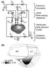
FIGURE 1
FIGURE 1
(a) Electric analog schematic of the circulatory model with the ventricular finite element model embedded. Cap compliance of pulmonic arteries and capillaries; Cvp compliance of pulmonic veins; Cas compliance of systemic arteries and capillaries; Cvs compliance of systemic veins; Rap resistance of pulmonic arteries and capillaries; Rvp resistance of pulmonic veins; Ras resistance of systemic arteries and capillaries; Rvs resistance of systemic veins; Ela left atrial time-varying elastance; Era right atrial time-varying elastance; Rmitral mitral valve resistance; Rtricus tricuspid valve resistance; Rao aortic valve impedance; Rpa pulmonic artery impedance. Note that parts (e.g. pulmonic veins) of the circulation are lumped together and thus compliances are averaged and resistances summed over these parts. (b) Location of transmural ischemic region in the ventricular finite element model, shown on the endocardium. Parameter Ca0 represents the value of the intracellular calcium concentration bound to troponin C.
FIGURE 2
FIGURE 2
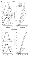
Static pressure and volume relations for a finite element (FE) model, decoupled from the circulation, and first-order fitted varying elastance (VE) model of the passive (a) and fully activated (b) canine heart. In 4 phases (numbers 1–4), pressures were prescribed in the ventricles, yielding cavity volumes for the left and right ventricles. Since the passive material used is elastic, it is time-independent. Therefore, we refer to these steps as load steps, since any time increment can be employed. The active material is kept maximally activated and thus is also time-independent at maximum activation. In phase 1, LV pressure (LVP) is increased and RV pressure (RVP) is kept constant. In phase 2, RVP is increased and LVP is constant. In phase 3, LVP is decreased while keeping RVP constant; and in phase 4, RVP is decreased while keeping LVP constant. Notice how the volume of the ventricle changes when pressure is kept at a constant level: a consequence of direct ventricular interaction. In the pressure–volume relation this shows up as horizontal plateaus. The alternating of increasing and decreasing LV and RV pressures, together with direct interaction, results in the observed PV loops.
FIGURE 3
FIGURE 3
Detailed flow chart of the complete pressure update algorithm. (a) Pressure update module; (b) modules of the circulatory model, perturbation of circulatory model, and FE model.
FIGURE 4
FIGURE 4
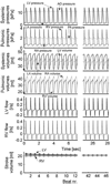
Hemodynamics for the first 12 and last 6 beats out of 46 from the NORM simulation. AO, aorta; LA, left atrium; LV, left ventricle; PA, pulmonary artery; RA, right atrium; RV, right ventricle. Note that aortic pressure is in fact mean pressure over the aorta, arteries and capillaries. The same applies for pulmonary artery pressure.
FIGURE 5
FIGURE 5
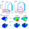
LV and RV PV loops for the (a) ISCH and (b) PAC simulation. For comparison, PV loops for the NORM simulation are also shown. (c) Endocardial myofiber strains in the deformed LV and RV at end-systole from the NORM (left column), ISCH, and PAC simulations. The black arrow points at larger strains in the ischemic region. The white arrow points at the septum, which is shifted towards the LV.
FIGURE 6
FIGURE 6
Hemodynamics for the (a) ISCH, and (b) PAC simulation. The first beat shown is the 12th beat of the normal heart. For abbreviations see Fig. 4.

References

    1. Bovendeerd PHM, Arts T, Delhaas T, Huyghe JM, Vancampen DH, Reneman RS. Regional wall mechanics in the ischemic left ventricle: Numerical modeling and dog experiments. Am. J. Physiol. Heart Circ. Physiol. 1996;39:H398–H410. - PubMed
    1. Bovendeerd PHM, Arts T, Huyghe JM, van Campen DH, Reneman RS. Dependence of local left-ventricular wall mechanics on myocardial fiber orientation – a model study. J. Biomech. 1992;25:1129–1140. - PubMed
    1. Brickner ME, Hillis LD. Congenital heart disease in adults – first of two parts. N. Engl. J. Med. 2000;342:256–263. - PubMed
    1. Burkhoff D, de Tombe PP, Hunter WC. Impact of ejection on magnitude and time course of ventricular pressure-generating capacity. Am. J. Physiol. Heart Circ. Physiol. 1993;265:H899–H909. - PubMed
    1. Carmeliet E. Cardiac ionic currents and acute ischemia: From channels to arrhythmias. Physiol. Rev. 1999;79:917–1017. - PubMed

Publication types