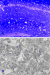
Spatial relationship between synapse loss and beta-amyloid deposition in Tg2576 mice
- PMID: 17111375
- PMCID: PMC1661843
- DOI: 10.1002/cne.21176
Spatial relationship between synapse loss and beta-amyloid deposition in Tg2576 mice
Abstract
Although there is evidence that beta-amyloid impairs synaptic function, the relationship between beta-amyloid and synapse loss is not well understood. In this study we assessed synapse density within the hippocampus and the entorhinal cortex of Tg2576 mice at 6-18 months of age using stereological methods at both the light and electron microscope levels. Under light microscopy we failed to find overall decreases in the density of synaptophysin-positive boutons in any brain areas selected, but bouton density was significantly decreased within 200 mum of compact beta-amyloid plaques in the outer molecular layer of the dentate gyrus and Layers II and III of the entorhinal cortex at 15-18 months of age in Tg 2576 mice. Under electron microscopy, we found overall decreases in synapse density in the outer molecular layer of the dentate gyrus at both 6-9 and 15-18 months of age, and in Layers II and III of the entorhinal cortex at 15-18 months of age in Tg 2576 mice. However, we did not find overall changes in synapse density in the stratum radiatum of the CA1 subfield. Furthermore, in the two former brain areas we found a correlation between lower synapse density and greater proximity to beta-amyloid plaques. These results provide the first quantitative morphological evidence at the ultrastructure level of a spatial relationship between beta-amyloid plaques and synapse loss within the hippocampus and the entorhinal cortex of Tg2576 mice.
Figures


References
-
- Albin RL, Greenamyre JT. Alternative excitotoxic hypotheses. Neurology. 1992;42:733–738. - PubMed
-
- Bannerman DM, Yee BK, Lemaire M, Wilbrecht L, Jarrard L, Iversen SD, Rawlins JN, Good MA. The role of the entorhinal cortex in two forms of spatial learning and memory. Exp Brain Res. 2001;141:281–303. - PubMed
-
- Barnes P, Good M. Impaired Pavlovian cued fear conditioning in Tg2576 mice expressing a human mutant amyloid precursor protein gene. Behav Brain Res. 2005;157:107–17. - PubMed
-
- Boncristiano S, Calhoun ME, Howard V, Bondolfi L, Kaeser SA, Wiederhold KH, Staufenbiel M, Jucker M. Neocortical synaptic bouton number is maintained despite robust amyloid deposition in APP23 transgenic mice. Neurobiol Aging. 2005;26:607–13. - PubMed
Publication types
MeSH terms
Substances
Grants and funding
LinkOut - more resources
Full Text Sources
Other Literature Sources
Medical
Miscellaneous

