. 2006 Nov;2(11):e124.
doi: 10.1371/journal.ppat.0020124.

Epitope mapping and topographic analysis of VAR2CSA DBL3X involved in P. falciparum placental sequestration

Affiliations

Epitope mapping and topographic analysis of VAR2CSA DBL3X involved in P. falciparum placental sequestration

Madeleine Dahlbäck et al. PLoS Pathog. 2006 Nov.

Abstract

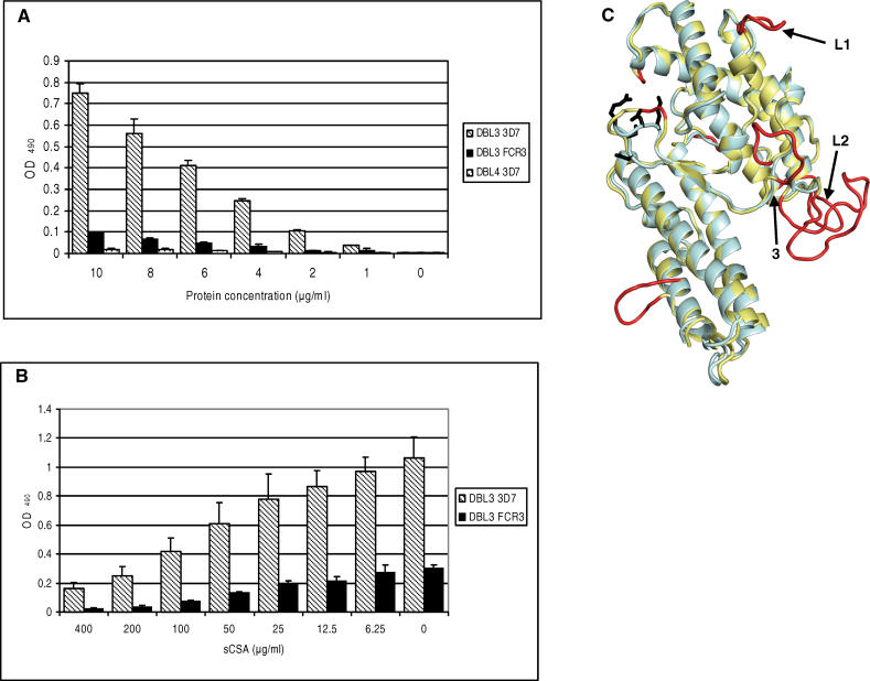
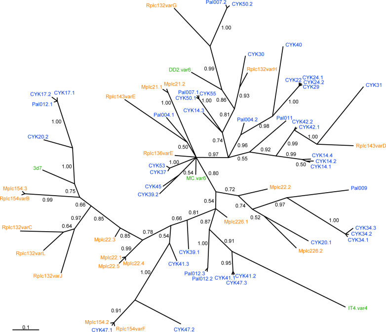
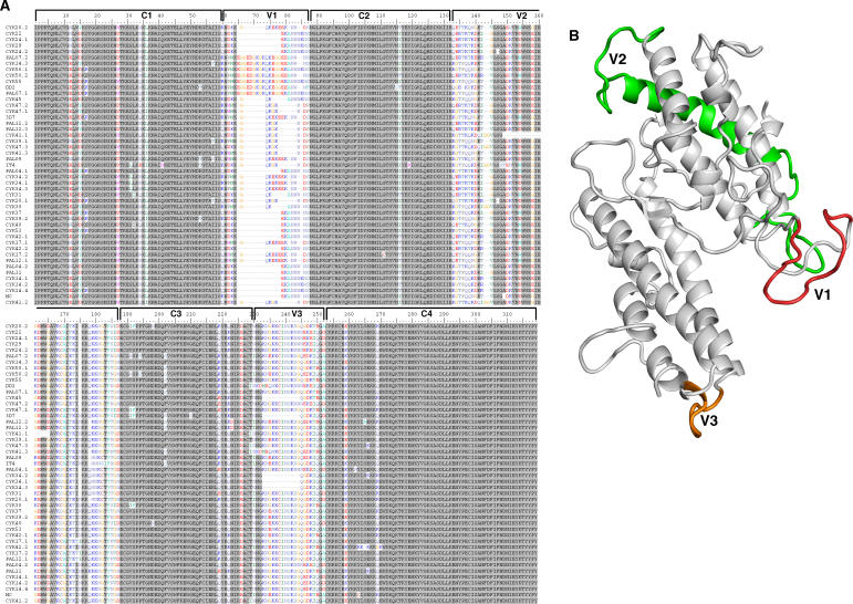
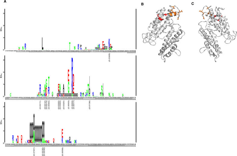
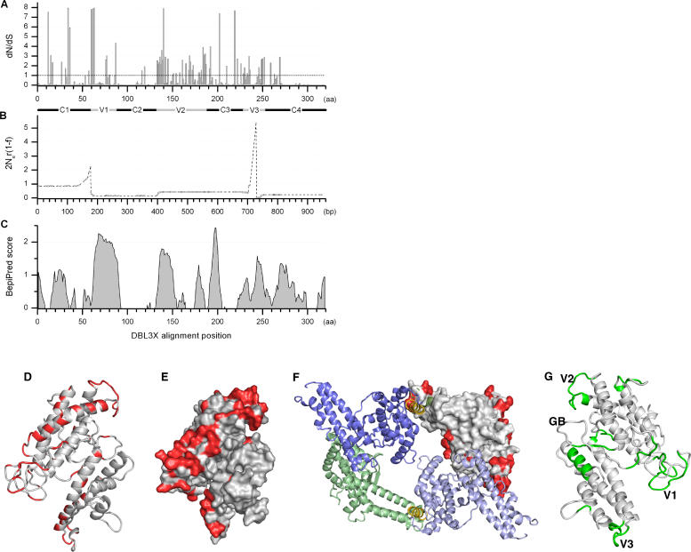
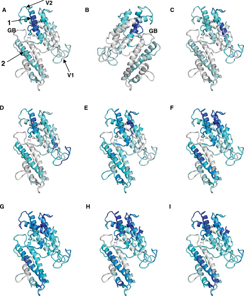
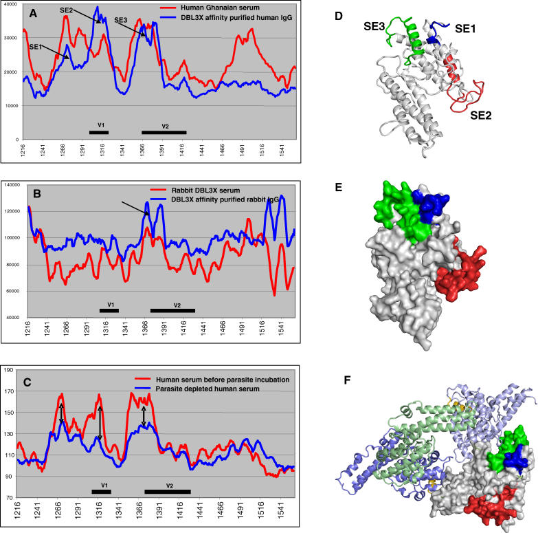
Pregnancy-associated malaria is a major health problem, which mainly affects primigravidae living in malaria endemic areas. The syndrome is precipitated by accumulation of infected erythrocytes in placental tissue through an interaction between chondroitin sulphate A on syncytiotrophoblasts and a parasite-encoded protein on the surface of infected erythrocytes, believed to be VAR2CSA. VAR2CSA is a polymorphic protein of approximately 3,000 amino acids forming six Duffy-binding-like (DBL) domains. For vaccine development it is important to define the antigenic targets for protective antibodies and to characterize the consequences of sequence variation. In this study, we used a combination of in silico tools, peptide arrays, and structural modeling to show that sequence variation mainly occurs in regions under strong diversifying selection, predicted to form flexible loops. These regions are the main targets of naturally acquired immunoglobulin gamma and accessible for antibodies reacting with native VAR2CSA on infected erythrocytes. Interestingly, surface reactive anti-VAR2CSA antibodies also target a conserved DBL3X region predicted to form an alpha-helix. Finally, we could identify DBL3X sequence motifs that were more likely to occur in parasites isolated from primi- and multigravidae, respectively. These findings strengthen the vaccine candidacy of VAR2CSA and will be important for choosing epitopes and variants of DBL3X to be included in a vaccine protecting women against pregnancy-associated malaria.


Conflict of interest statement

Competing interests. The authors have declared that no competing interests exist.

Figures

Figure 1
Figure 1. Recombinant DBL3X VAR2CSA Binds CSA and the Structure of the Domain Can Be Modeled on the Basis of the Structure of the DBL Domains of EBA-175
(A) Binding assay. DBL3X binds to CSA in a protein concentration-specific manner. ELISA plates were coated with soluble CSA and binding of recombinant proteins at different concentrations was determined. DBL3X from 3D7 shows better binding compared to DBL3X from FCR3. The non-CSA binding VAR2CSA DBL4ɛ was included as a control. Results are the mean of three binding assays and the error bars indicate the standard deviation. (B) Inhibition assay. Recombinant DBL3X proteins (7 μg/ml) were preincubated with increasing amounts of soluble CSA, and binding to CSA-coated plates was determined. Binding of DBL3X is dose-dependently inhibited by soluble CSA. Results are the mean of four inhibition binding assays and error bars indicate the standard deviation. (C) Model of DBL3X using EBA-175 as template. The figure shows a superposition of the EBA-175 F1 domain in light blue and the DBL3X (PFL0030c amino acids 1,217–1,559) model in yellow with insertions highlighted in red. Arrows indicate insertions, which align with flexible loop regions in Pkα-DBL (L1) and EBA-175 F1 (L2). Side chains of glycan-binding residues in EBA-175 F1 are shown in black. Arrow 3 indicates the corresponding Pkα-DBL Duffy antigen receptor for chemokine-binding site.
Figure 2
Figure 2. Phylogenetic Relationship between Different VAR2CSA DBL3X Sequences
Bayesian inference tree of 43 placental cDNA sequences from Senegal (blue), 21 Malawian sequences (orange), and database sequences from four isolates (green) of different geographic origin. The posterior probability of the clades is shown at the branches.
Figure 3
Figure 3. Multiple Alignment of VAR2CSA DBL3X Sequences
(A) cDNA from 43 placental parasite samples were amplified with conserved DBL3X primers and sequenced. Sequences were curated for primer sequence, translated, and aligned. The text to the left indicates the identity of the samples. The DBL3X domain can be divided into four regions, which are highly conserved, C1–C4; and three regions, which are polymorphic and harbor deletions V1–V3. (B) Model of DBL3X showing the position of V1 (red), V2 (green), and V3 (orange).
Figure 4
Figure 4. Sequence Differences in Infections with Different Parity
(A) Kullback-Leibler sequence logo based on a multiple alignment of the DBL3X region (Figure 3). The sequences in the alignment were split into two groups of equal size: 21 sequences obtained from primigravidae and 21 sequences from multigravidae women. The x-axis shows amino acid positions in the alignment. The height of a position indicates the amount of difference between the two groups according to the symmetric Kullback-Leibler distance, and the letters indicate the amino acids contributing to this difference. The letter “O” has been chosen to signify gaps in the alignment. Polar amino acids are green, basic are blue, acidic are red, and hydrophobic amino acids are black. P-values indicating the significance of DKL based on the parity grouping compared to random groupings are shown below positions where p < 0.05. (B) DBL3X model showing the position of amino acids that were differentially found in primi-gravidae and multigravidae women. Residues in the region 135–175 of moderately significant difference are shown in orange, and highly significant residues (positions 158, 161, and 162) are shown in red. (C) The model (B) rotated 180 degrees so it is positioned similar to Figure 1C.
Figure 5
Figure 5. Sequence Variation and B Cell Epitopes in DBL3
(A) dN/dS ratio per amino acid based on an alignment of 47 DBL3X sequences. dN/dS ratio higher than the dashed line indicates that the position is under positive selection. V1–V3 and C1–C4 are indicated with solid bars. It should be noted that the inference of selection pressure is less reliable in positions with gaps, which are mainly found in the variable regions (see Figure 3). (B) The population recombination rate ρ = 2Ner(1 − f) estimated per bp across DBL3X. Circles indicate the SNPs for which ρ is estimated, linear interpolation is shown between SNPs. Two hotspots corresponding to a local raise in r are observed in association with the variable regions V1 and V3. (C) B cell epitope predictions of the 3D7 sequence performed by the BepiPred server. DD2 and IT-4 were used to fill 3D7 sequence gaps. (D) Cartoon representation of the DBL3X model showing amino acid positions under diversifying selection in red (dN/dS ratios >1). (E) DBL3X model with surface topography prediction showing amino acid positions under diversifying selection in red (dN/dS ratios >1). (F) The homo-dimer of the two domains of RII of EBA-175 has been shown to form a handshake structure with F1 of one molecule dimerizing with F2 of another molecule and vice-versa. Here VAR2CSA DBL3X has been superimposed on the F1 domain of one of the EBA-175 molecules in the dimer. The F1 domain is shown in green, F2 domains are blue, and the linker regions are gold. Amino acids under diversifying selection in DBL3X are shown in red (dN/dS ratios >1). It is clear that most residues inward of the dimer are not under positive selection. (G) DBL3X model showing residues with BepiPred scores higher than 0.9 in green. Locations of V1–V3 are indicated on the model. GB indicates the glycan-binding site of EBA-175.
Figure 6
Figure 6. Defining Targets of Antibodies on DBL3X
Models of DBL3X in which the intensity of the gray and blue indicates reactivity of plasma tested on a peptide array (PepScan scores). Gray indicates lowest scores and dark blue indicates highest scores. (A and C–I) represent reactivity in eight individual plasma samples. (B) shows the reactivity in the same plasma as tested in (A), but the model has been rotated 180 degrees. Arrow 1 indicates a highly recognized conserved α-helix region, which was not predicted to be an epitope by BepiPred and was characterized by low dN/dS ratios and low sequence variation. Arrow 2 indicates another α-helix with a predicted B cell epitope containing some residues with high sequence variation. Variable regions V1 and V2 are fairly well recognized by most of the serum samples. GB indicates the glycan-binding site of EBA-175.
Figure 7
Figure 7. Reactivity of Plasma Affinity Purified on Recombinant DBL3X and Human IgG Depleted for Surface Reactivity by Incubation with Infected Erythrocytes Expressing VAR2CSA
(A) PepScan IgG reactivity in a Ghanaian pool of pregnancy plasma before (red) and after (blue) affinity purification on recombinant DBL3X protein. Variable regions V1 and V2 are indicated with a black line. Arrows indicate three surface-exposed regions (SE). The x-axis is amino acid position in 3D7 VAR2CSA. (B) PepScan IgG reactivity in plasma from a rabbit immunized with DBL3X before (red) and after (blue) affinity purification on recombinant DBL3X protein. Arrow indicates the surface-exposed region. N- and C-terminal parts of the recombinant proteins did also appear to be surface-exposed; this could be due to improper folding of the N- and C-terminal parts of the protein. (C) PepScan IgG reactivity in a Tanzanian pool of pregnancy plasma before (red) and after (blue) depletion of antibodies directed against surfaced-exposed VAR2CSA regions by incubation with infected erythrocytes expressing VAR2CSA on the surface. Arrows indicate three regions where the reactivity was markedly reduced after depletion largely corresponding to SE1, SE2, and SE3 on (A). (D) DBL3X model showing the location of surface-exposed epitopes, SE1 (blue), SE2 (red), and SE3 (green). (E) DBL3X surface topography showing SE1–SE3. From the model it is apparent that the SE1 and SE3 form a continuous epitope. (F) VAR2CSA DBL3X superimposed on the F1 domain of one of the molecules in the EBA-175 dimer. The F1 domain is shown in green, F2 domains are blue, and the linker regions are gold. On the DBL3X domain the three experimentally verified surface-exposed regions are shown in similar coloring as (D and E) The three regions are mainly predicted to be on the opposite side of the central cavity of the dimer, and parts of SE3 correspond to amino acid positions directly involved in the dimerization of EBA-175.

Similar articles

Cited by

References

    1. Christophers SR. The mechanism of immunity against malaria in communities living under hyper-endemic conditions. Ind J Med Res. 1924;12:273–294.
    1. Brabin BJ. An analysis of malaria in pregnancy in Africa. Bull World Health Organ. 1983;61:1005–1016. - PMC - PubMed
    1. Walter PR, Garin Y, Blot P. Placental pathologic changes in malaria. A histologic and ultrastructural study. Am J Pathol. 1982;109:330–342. - PMC - PubMed
    1. Bray RS, Anderson MJ. Falciparum malaria and pregnancy. Trans R Soc Trop Med Hyg. 1979;73:427–431. - PubMed
    1. McGregor IA, Wilson ME, Billewicz WZ. Malaria infection of the placenta in Gambia, West Africa: Its incidence and relationship to stillbirth, birth-weight, and placental weight. Trans R Soc Trop Med Hyg. 1983;77:232–244. - PubMed

Publication types

MeSH terms

Substances

Associated data