

Myelination and nodal formation of regenerated peripheral nerve fibers following transplantation of acutely prepared olfactory ensheathing cells
- PMID: 17112480
- PMCID: PMC2673087
- DOI: 10.1016/j.brainres.2006.09.089
Myelination and nodal formation of regenerated peripheral nerve fibers following transplantation of acutely prepared olfactory ensheathing cells
Abstract
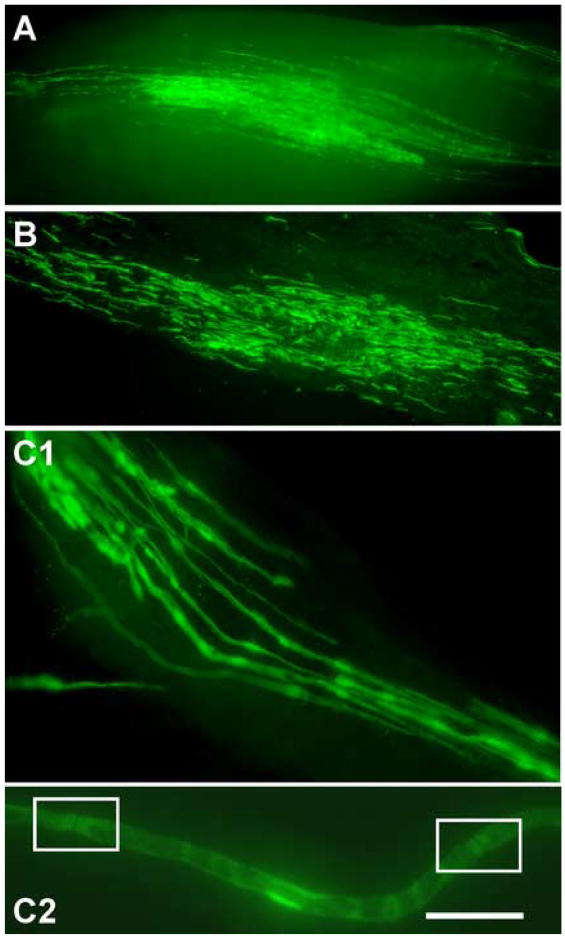
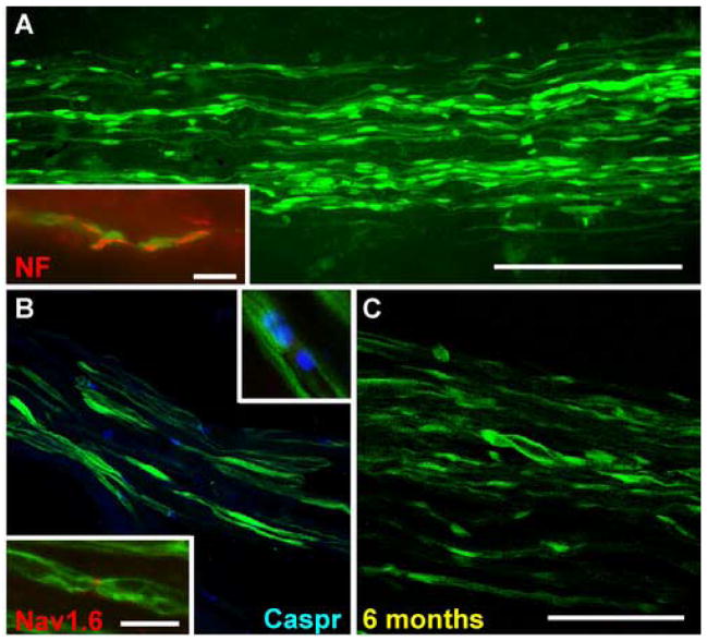
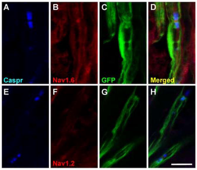
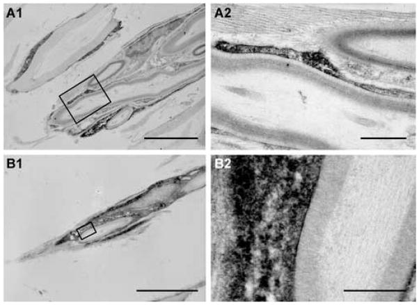
Transplantation of olfactory ensheathing cells (OECs) into injured spinal cord results in improved functional outcome. Mechanisms suggested to account for this functional improvement include axonal regeneration, remyelination and neuroprotection. OECs transplanted into transected peripheral nerve have been shown to modify peripheral axonal regeneration and functional outcome. However, little is known of the detailed integration of OECs at the transplantation site in peripheral nerve. To address this issue, cell populations enriched in OECs were isolated from the olfactory bulbs of adult green fluorescent protein (GFP)-expressing transgenic rats and transplanted into a sciatic nerve crush lesion which transects all axons. Five weeks to 6 months after transplantation, the nerves were studied histologically. GFP-expressing OECs survived in the lesion and distributed longitudinally across the lesion zone. The internodal regions of individual teased fibers distal to the transection site were characterized by GFP expression in the cytoplasmic and nuclear compartments of cells surrounding the axons. Immunoelectron microscopy for GFP indicated that the transplanted OECs formed peripheral type myelin. Immunostaining for sodium channel and Caspr revealed a high density of Na(v)1.6 at the newly formed nodes of Ranvier which were flanked by paranodal Caspr staining. These results indicate that transplanted OECs extensively integrate into transected peripheral nerve and form myelin on regenerated peripheral nerve fibers, and that nodes of Ranvier of these axons display proper sodium channel organization.
Figures
Similar articles
-
Integration of engrafted Schwann cells into injured peripheral nerve: axonal association and nodal formation on regenerated axons.Neurosci Lett. 2005 Oct 21;387(2):85-9. doi: 10.1016/j.neulet.2005.06.073. Neurosci Lett. 2005. PMID: 16084645 Free PMC article.
-
Transplantation of olfactory ensheathing cells enhances peripheral nerve regeneration after microsurgical nerve repair.Brain Res. 2009 Feb 13;1254:10-7. doi: 10.1016/j.brainres.2008.11.036. Epub 2008 Nov 21. Brain Res. 2009. PMID: 19059220
-
Molecular reconstruction of nodes of Ranvier after remyelination by transplanted olfactory ensheathing cells in the demyelinated spinal cord.J Neurosci. 2006 Feb 8;26(6):1803-12. doi: 10.1523/JNEUROSCI.3611-05.2006. J Neurosci. 2006. PMID: 16467529 Free PMC article.
-
[New insights on the organization of the nodes of Ranvier].Rev Neurol (Paris). 2014 Dec;170(12):819-24. doi: 10.1016/j.neurol.2014.03.017. Epub 2014 Nov 20. Rev Neurol (Paris). 2014. PMID: 25459119 Review. French.
-
Molecular organization and function of vertebrate septate-like junctions.Biochim Biophys Acta Biomembr. 2020 May 1;1862(5):183211. doi: 10.1016/j.bbamem.2020.183211. Epub 2020 Feb 4. Biochim Biophys Acta Biomembr. 2020. PMID: 32032590 Review.
Cited by
-
The Effect of Schwann Cells/Schwann Cell-Like Cells on Cell Therapy for Peripheral Neuropathy.Front Cell Neurosci. 2022 Mar 8;16:836931. doi: 10.3389/fncel.2022.836931. eCollection 2022. Front Cell Neurosci. 2022. PMID: 35350167 Free PMC article. Review.
-
Peripheral Nerve Regeneration Using a Nerve Conduit with Olfactory Ensheathing Cells in a Rat Model.Tissue Eng Regen Med. 2021 Jun;18(3):453-465. doi: 10.1007/s13770-020-00326-9. Epub 2021 Jan 30. Tissue Eng Regen Med. 2021. PMID: 33515167 Free PMC article.
-
Peripheral nerve regeneration: a current perspective.Eplasty. 2009 Oct 12;9:e47. Eplasty. 2009. PMID: 19907643 Free PMC article.
-
Demyelinating diseases and potential repair strategies.Int J Dev Neurosci. 2007 May;25(3):149-53. doi: 10.1016/j.ijdevneu.2007.02.002. Epub 2007 Mar 3. Int J Dev Neurosci. 2007. PMID: 17408905 Free PMC article. Review.
-
Peripheral nerve injuries and transplantation of olfactory ensheathing cells for axonal regeneration and remyelination: fact or fiction?Int J Mol Sci. 2012 Oct 10;13(10):12911-24. doi: 10.3390/ijms131012911. Int J Mol Sci. 2012. PMID: 23202929 Free PMC article. Review.
References
-
- Bareyre FM, Kerschensteiner M, Raineteau O, Mettenleiter TC, Weinmann O, Schwab ME. The injured spinal cord spontaneously forms a new intraspinal circuit in adult rats. Nat Neurosci. 2004;10:269–277. - PubMed
-
- Black JA, Cummins TR, Plumpton C, Chen YH, Hormuzdiar W, Clare JJ, Waxman SG. Upregulation of a silent sodium channel after peripheral, but not central nerve injury in DRG neurons. J Neurophysiol. 1999;82:2776–2785. - PubMed
-
- Black JA, Renganathan M, Waxman SG. Sodium channel Na (v) 1.6 is expressed along nonmyelinated axons and it contributes to conduction. Brain Res. Mol Brain Res. 2002;105(1–2):19–28. - PubMed
Publication types
MeSH terms
Substances
Grants and funding
LinkOut - more resources
Full Text Sources
Other Literature Sources
