. 2007 Jan 19;555(1):67-75.
doi: 10.1016/j.ejphar.2006.10.027. Epub 2006 Oct 19.

Serotonin in microdialysate from the mediobasal hypothalamus increases after progesterone administration to estrogen primed macaques

Affiliations

Serotonin in microdialysate from the mediobasal hypothalamus increases after progesterone administration to estrogen primed macaques

Maria Luisa Centeno et al. Eur J Pharmacol. .

Abstract

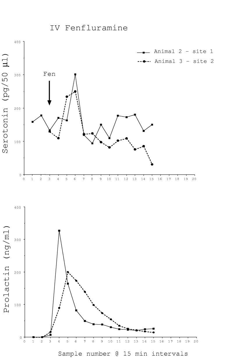
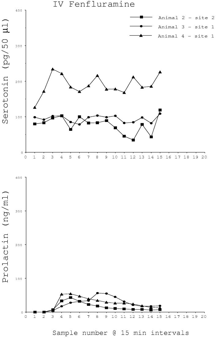
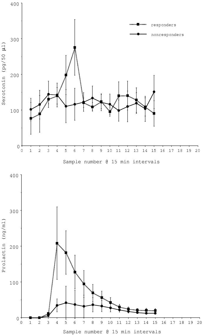
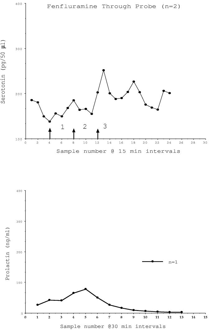
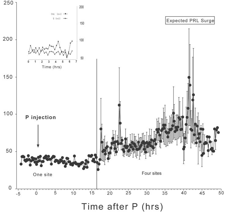
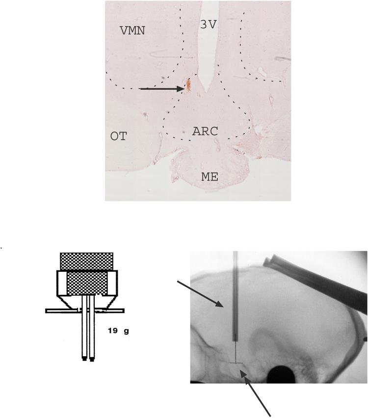
Estrogen and progesterone act on gene and protein expression in serotonin neurons in a manner that suggests serotonin neurotransmission should increase. However, measurement of extracellular serotonin in macaques was lacking. Elevated prolactin secretion can be an indicator of increased serotonergic function and prolactin is increased by combined estrogen and progesterone treatment. We examined extracellular serotonin by microdialysis in a well-characterized macaque model of steroid-induced prolactin secretion. Monkeys were fitted with 2 guide tubes directed to the arcuate nucleus of the hypothalamus. Samples (75 microl/15-minute interval) were obtained via a tether-swivel device through sample lines into an adjoining room. Serotonin was measured with a modified commercial enzyme linked immunoassay (ELISA) kit. Fenfluramine infused through the probe (300 microM for 2 h; n=2 trials) or administered intravenously (2.5 mg/kg; n=2 trials) caused a marked increase in extracellular serotonin and verified the efficacy of the procedure. Three monkeys were maintained with an estrogen implant for 2 weeks. Each monkey was injected with 20 mg of progesterone s.c. in oil at 1500 h; microdialysis was initiated the next morning and samples were obtained for 24 h. There was a significant increase in serotonin between 40 and 43 h after the progesterone injection (P<0.001, ANOVA). Serotonin averaged 59+/-1 pg/sample from 18-30 h post-progesterone injection, and averaged 76+/-2 pg/sample from 30-48 h post-progesterone injection (P<0.0001; t-test). Since the increase in serotonin is delayed by approximately 40 h after progesterone-injection, we speculate that the action of progesterone may involve either nuclear progestin receptors or membrane progestin receptors.


Figures

Fig. 1A
Fig. 1A
Microdialysate serotonin (pg/50μl) and serum prolactin (ng/ml) concentrations in two monkeys who responded robustly to intravenous administration of fenfluramine (2.5 mg/kg). With the time required for the serotonin in the brain to reach the sample tube equaling approximately 1 hour, the increase in serotonin and the increase in prolactin are temporally correlated.
Fig. 1B
Fig. 1B
Microdialysate serotonin (pg/50μl) and serum prolactin (ng/ml) concentrations in 3 monkeys who did not respond to intravenous administration of fenfluramine (2.5 mg/kg). There was no apparent increase in extracellular serotonin although prolactin exhibited a modest increase.
Fig. 1C
Fig. 1C
Average serotonin and prolactin concentrations in i.v. fenfluramine responders (n=2) and non-responders (n=3). There was a near significant difference in serotonin between the groups (P < 0.05; 2 way ANOVA) and a highly significant difference in prolactin between the groups (P < 0.0001; 2 way ANOVA).
Fig. 2
Fig. 2
Microdialysate serotonin (pg/50μl) and serum prolactin (ng/ml) concentrations in animals that were administered fenfluramine through the microdialysis probe. In 2 monkeys, an increase in extracellular serotonin was detected at the time expected based upon a traverse time to the brain of 1 hour and a traverse time from the brain to the sample tube of 1 hour. An increase in serum prolactin was detected at the time when fenfluramine reached the brain in one animal with a patent venous catheter. The arrows demarcate (1) the time at which the fenfluramine buffer was initiated, (2) the time at which the fenfluramine reached the brain and (3) the time that the potential increase in serotonin could first reach a sample tube.
Fig. 3
Fig. 3
Microdialysate serotonin (pg/50μl) from 18 to 48 h at 15-minute intervals after an injection of progesterone to estrogen-primed animals (n=4). In one animal, samples were collected prior to and immediately following progesterone injection, as well. Microdialysate concentrations of serotonin were elevated over baseline from 40-43 h post progesterone injections and correlate temporally with progesterone-induced prolactin secretion from earlier studies. The overall concentration of serotonin is significantly higher between 30 and 48 h than between 15 and 30 h post progesterone injection (P < 0.001; t-test). The average serotonin concentrations of the 4 sites were further parsed into time brackets containing 3 means per bracket from 17.5 to 48 h post progesterone injection, which yielded 41 time brackets. There was a significant difference in serotonin across time (P < 0.0001, one way ANOVA). Time brackets 32, 33, and 34 corresponding to 40.75 to 42.75 h post progesterone injection were significantly different from all other time brackets (P < 0.05, Student-Newman-Keuls posthoc pairwise comparison). Insert. Microdialysate serotonin (pg/50μl) from 0-7 h at 15-minute intervals in an untreated ovariectomized monkey and in 2 monkeys primed with estrogen. Concentrations of serotonin remained low during this period.
Fig. 4
Fig. 4
Montage of 10x images of the mediobasal hypothalamus of one representative monkey showing the posterior probe placement with DAB staining. The DAB injection site is located near the bottom of the 3rd ventricle of the mediobasal hypothalamus. The anterior site would have been located 2 mm in front of this point. In addition, the design of the headpiece holding the guide cannulae, and a lateral roentgenogram from the same animal (#4), are shown. The xray was obtained at final cannula placement and indicates the cannulae barrels (single-headed arrow). The double-headed arrow indicates the pencil mark on film, which marks the midpoint of the edges of the sella turcica. The single arrowheads indicate the anterior and posterior edges of the sella turcica, which are the critical landmarks for positioning of the guide cannulae. The guide cannulae are positioned at equal distance from the anterior and posterior points. The microdialysis probe extends several millimeters beyond the end of the guide tubes. All of the data reported for the estrogen+progesterone experiments are from animals with similar probe locations. This location contains arcuate nucleus neurons. Abbreviations: ARC-arcuate nucleus; ME- median eminence; OT- optic track; VMN- ventromedial nucleus; 3V- third ventricle.

References

    1. Azmitia EC. The serotonin-producing neurons of the midbrain median and dorsal raphe nuclei. In: Iverson LL, Iverson SD, Synder SH, editors. Handbook of Psychopharmacology. Vol. 9. Plenum Publishing Corp.; Baltimore, MD: 1978. pp. 233–313.
    1. Azmitia EC, Gannon PJ. The primate serotonergic system: a review of human and animal studies and a report on macaca fascicularis. Adv. Neurol. 1986;43:407–468. - PubMed
    1. Azmitia EC, Segal M. An autoradiographic analysis of the differential ascending projections of the dorsal raphe and median raphe nuclei in the rat. J. Comp. Neurol. 1978;179:641–668. - PubMed
    1. Bethea CL. Colocalization of progestin receptors with serotonin in raphe neurons of macaque. Neuroendocrinology. 1993;57:1–6. - PubMed
    1. Bethea CL. Regulation of progestin receptors in raphe neurons of steroid-treated monkeys. Neuroendocrinology. 1994;60:50–61. - PubMed

Publication types