. 2007 Mar 30;360(1):6-16.
doi: 10.1016/j.virol.2006.09.017. Epub 2006 Nov 17.

Bone-marrow-derived mesenchymal stem cells as a target for cytomegalovirus infection: implications for hematopoiesis, self-renewal and differentiation potential

Affiliations

Bone-marrow-derived mesenchymal stem cells as a target for cytomegalovirus infection: implications for hematopoiesis, self-renewal and differentiation potential

Sergey V Smirnov et al. Virology. .

Abstract

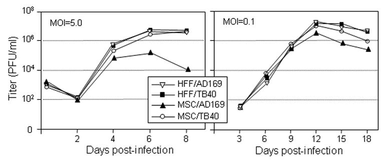
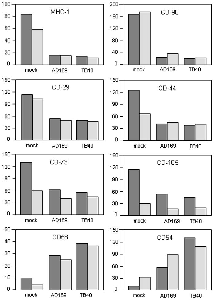
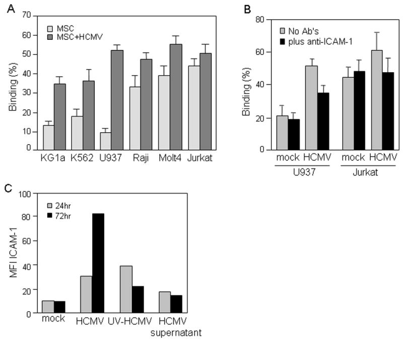
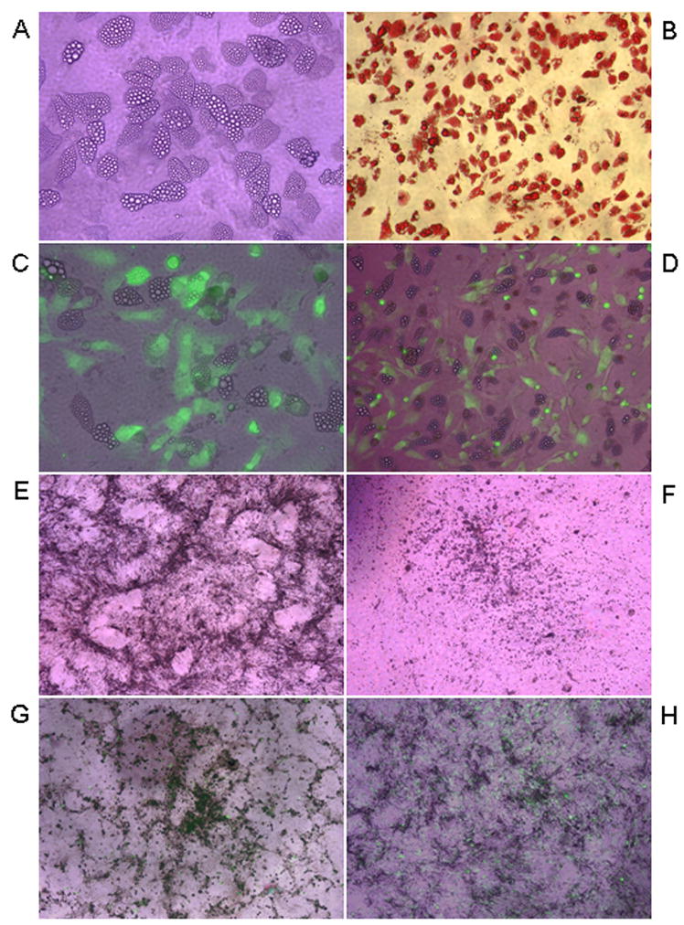
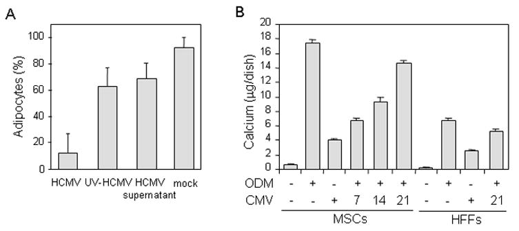
Mesenchymal stem cells (MSCs) in bone marrow (BM) regulate the differentiation and proliferation of adjacent hematopoietic precursor cells and contribute to the regeneration of mesenchymal tissues, including bone, cartilage, fat and connective tissue. BM is an important site for the pathogenesis of human cytomegalovirus (HCMV) where the virus establishes latency in hematopoietic progenitors and can transmit after reactivation to neighboring cells. Here we demonstrate that BM-MSCs are permissive to productive HCMV infection, and that HCMV alters the function of MSCs: (i) by changing the repertoire of cell surface molecules in BM-MSCs, HCMV modifies the pattern of interaction between BM-MSCs and hematopoietic cells; (ii) HCMV infection of BM-MSCs undergoing adipogenic or osteogenic differentiation impaired the process of differentiation. Our results suggest that by altering BM-MSC biology, HCMV may contribute to the development of various diseases.


Figures

Fig. 1
Fig. 1. Characterization of HCMV infection in MSCs
Growth kinetics of laboratory AD169 and clinical TB40 HCMV strains were compared in MSC and HFF cells. The cells were infected with virus at a high MOI of 5 PFU/cell (single-step growth curves, left panel) or at a low MOI of 0.1 PFU/cell (multistep growth curves, right pane). Cell-free and cell-associated virus was collected at indicated times and the virus titer was determined. Shown data is from one representative experiment out of three independent experiments.
Fig. 2
Fig. 2. Modulation of cell surface molecule expression in MSC and HFF cells infected with HCMV
Analysis of the expression of cell surface molecules on MSCs (dark shaded bars) and HFFs (light shaded bars) was performed with the use of flow cytometry. Uninfected or HCMV-infected (3 days post infection) cells were collected and incubated with the following conjugated monoclonal antibodies: CD29-PE, CD44-PE, CD73-PE, CD90-PE, CD105-FITC and HLA-ABC-PE; or with CD54 or CD58 antibodies and subsequently with FITC-conjugated secondary antibody. Nonspecific fluorescence was determined by incubation of the cells with isotype-matched mouse monoclonal antibodies. Antibody-treated cells were subjected to flow cytometry. Data are expressed as relative fluorescence intensity (RFI) as a percentage of the baseline value (isotype control). Shown data is from one representative experiment out of three independent experiments.
Fig. 3
Fig. 3. Effect of HCMV infection on interaction between MSCs and hematopoietic cells
(A and B) Adherent MSCs were seeded in 96-well plates, left uninfected or infected with TB40 HCMV at MOI of 3–5 PFU/cell for 72 hr, and treated with blocking ICAM antibody (B) or left untreated (A). Suspension cell lines shown on the figure were labeled with PKH26 red fluorescent dye and allowed to adhere to MSCs for 1 hr at 37°C at a ratio of 5–10 suspension/monolayer cells. Unbound cells were washed out and the fluorescence of cells retained in the wells was measured. Results are expressed as the percentage of adherent cells. The adhesion assay was performed in quadruplicate format for each target cell line. (C) MSCs were left uninfected (mock), infected with TB40 HCMV at MOI of 3–5 PFU/cell or treated with either UV-inactivated HCMV virions (UV-HCMV) or HCMV-free supernatants (HCMV supernatant); and ICAM-1 expression was analyzed by flow cytometry at 24 and 72 hr post infection. Data are expressed as RFI as a percentage of the baseline value (isotype control). Shown data is from one representative experiment out of three independent experiments.
Fig. 4
Fig. 4. HCMV infection of MSCs differentiating into adipogenic or osteogenic lineage
MSCs were incubated in either adipogenic (A–D) or osteogenic (E, G and H) differentiation media for 28 days, left uninfected or infected with TB40 BAC-GFP HCMV at MOI of 3–5 PFU/cell at days 0, 7, 14 and 21 of differentiation. HFFs (F) were also incubated in osteogenic differentiation media for 28 days. All cultures were analyzed at day 28 of the differentiation. Lipid vacuole accumulation in uninfected adipogenic MSC cultures was observed by regular light microscopy (A; X600, regular light microscopy) and was confirmed with Oil red O staining (B; X400, regular light microscopy). GFP expression was used to monitor productive HCMV infection in adipogenic cultures infected at day 14 (C; X600, merged regular and fluorescent light microscopy) or day 21 of differentiation (D; X400; merged regular and fluorescent light microscopy). MSCs (E) and HFFs (F), incubated in osteogenic differentiation media, were photographed to observe calcium deposition in the cells (X400; regular light microscopy). GFP expression was used to monitor productive HCMV infection in osteogenic cultures infected at day 7 (G) or day 21 of differentiation (H) (X400; merged regular and fluorescent light microscopy).
Fig. 5
Fig. 5. Effect of HCMV infection on adipogenesis and osteogenesis
(A) MSCs were seeded in 12 well plates and left uninfected (mock), infected with TB40 HCMV at MOI of 3–5 PFU/cell or treated with either UV-inactivated HCMV virions (UV-HCMV) or HCMV-free supernatants (HCMV supernatant) for 1 day at the beginning of each cycle of induction/maintenance and then maintained in differentiation medium on the same schedule as described above in figure 4 legend for 2 cycles. After 14 days the adipogenic cultures were stained with Oil red O and adipocytes (cells containing lipid vacuoles) were counted. The number of adipocytes is shown as a percentage of the total number of all cells. Data represents results of 5 counting’s in each out of three wells. (B) MSCs and HFFs were cultured in standard or osteogenic differentiation media (ODM) for 28 days. Cells were left uninfected or infected with TB40 BAC-GFP HCMV at indicated days (7, 14 and 21) of osteogenic differentiation. Calcium deposition in the cells was determined at day 28. Total calcium (μg/dish) was calculated from standard solutions. Assays were done in triplicate at each time point.

References

    1. Ahn K, Angulo A, Ghazal P, Peterson PA, Yang Y, Fruh K. Human cytomegalovirus inhibits antigen presentation by a sequential multistep process. Proc Natl Acad Sci USA. 1996;93 (20):10990–10995. - PMC - PubMed
    1. Apperley JF, Dowding C, Hibbin J, Buiter J, Matutes E, Sissons PJ, Gordon M, Goldman JM. The effect of cytomegalovirus on hemopoiesis: in vitro evidence for selective infection of marrow stromal cells. Exp Hematol. 1989;17 (1):38–45. - PubMed
    1. Baldick CJ, Jr, Marchini A, Patterson CE, Shenk T. Human cytomegalovirus tegument protein pp71 (ppUL82) enhances the infectivity of viral DNA and accelerates the infectious cycle. J Virol. 1997;71 (6):4400–4408. - PMC - PubMed
    1. Boeckh M, Nichols WG, Papanicolaou G, Rubin R, Wingard JR, Zaia J. Cytomegalovirus in hematopoietic stem cell transplant recipients: Current status, known challenges, and future strategies. Biol Blood Marrow Transplant. 2003;9 (9):543–558. - PubMed
    1. Bruder SP, Jaiswal N, Haynesworth SE. Growth kinetics, self-renewal, and the osteogenic potential of purified human mesenchymal stem cells during extensive subcultivation and following cryopreservation. J Cell Biochem. 1997;64 (2):278–294. - PubMed

Publication types

MeSH terms

Substances