

Lasting changes in neuronal activation patterns in select forebrain regions of aggressive, adolescent anabolic/androgenic steroid-treated hamsters
- PMID: 17113655
- PMCID: PMC1829410
- DOI: 10.1016/j.bbr.2006.10.025
Lasting changes in neuronal activation patterns in select forebrain regions of aggressive, adolescent anabolic/androgenic steroid-treated hamsters
Abstract
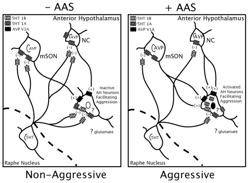
Repeated exposure to anabolic/androgenic steroids (AAS) during adolescence stimulates high levels of offensive aggression in Syrian hamsters. The current study investigated whether adolescent AAS exposure activated neurons in areas of hamster forebrain implicated in aggressive behavior by examining the expression of FOS, i.e., the protein product of the immediate early gene c-fos shown to be a reliably sensitive marker of neuronal activation. Adolescent AAS-treated hamsters and sesame oil-treated littermates were scored for offensive aggression and then sacrificed 1 day later and examined for the number of FOS immunoreactive (FOS-ir) cells in regions of the hamster forebrain important for aggression control. When compared with non-aggressive, oil-treated controls, aggressive AAS-treated hamsters showed persistent increases in the number of FOS-ir cells in select aggression regions, namely the anterior hypothalamus and lateral septum. However, no differences in FOS-ir cells were found in other areas implicated in aggression such as the ventrolateral hypothalamus, bed nucleus of the stria terminals, central and/or medial amygdala or in non-aggression areas, such as the samatosensory cortex and the suprachiasmatic nucleus. These results suggest that adolescent AAS exposure may constitutively activate neurons in select forebrain areas critical for the regulation of aggression in hamsters. A model for how persistent activation of neurons in one of these brain regions (i.e., the anterior hypothalamus) may facilitate the development of the aggressive phenotype in adolescent-AAS exposed animals is presented.
Figures


References
-
- Aoki C, Kaneko T, Starr A, Pickel VM. Identification of mitochondrial and non-mitochondrial glutaminase within select neurons and glia of rat forebrain by electron microscopic immunocytochemistry. J Neurosci Res. 1991;28:531–48. - PubMed
-
- Bamshad M, Albers HE. Neural circuitry controlling vasopressin-stimulated scent marking in Syrian hamsters (Mesocricetus auratus) J Comp Neurol. 1996;369:252–63. - PubMed
-
- Bamshad M, Cooper TT, Karom M, Albers HE. Glutamate and vasopressin interact to control scent marking in Syrian hamsters (Mesocricetus auratus) Brain Res. 1996;731:213–6. - PubMed
-
- Bamshad M, Karom M, Pallier P, Albers HE. Role of the central amygdala in social communication in Syrian hamsters (Mesocricetus auratus) Brain Res. 1997;744:15–22. - PubMed
-
- Brann DW, Mahesh VB. Glutamate: a major neuroendocrine excitatory signal mediating steroid effects on gonadotropin secretion. J Steroid Biochem Mol Biol. 1995;53:325–9. - PubMed
Publication types
MeSH terms
Substances
Grants and funding
LinkOut - more resources
Full Text Sources
Medical
