

The role of estrogen in the initiation of breast cancer
- PMID: 17113977
- PMCID: PMC1832080
- DOI: 10.1016/j.jsbmb.2006.09.004
The role of estrogen in the initiation of breast cancer
Abstract
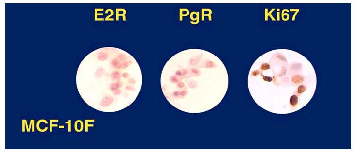
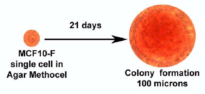
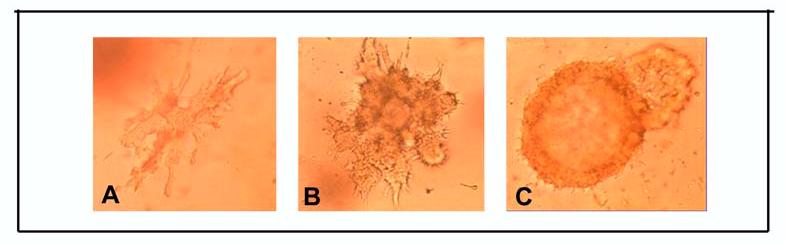
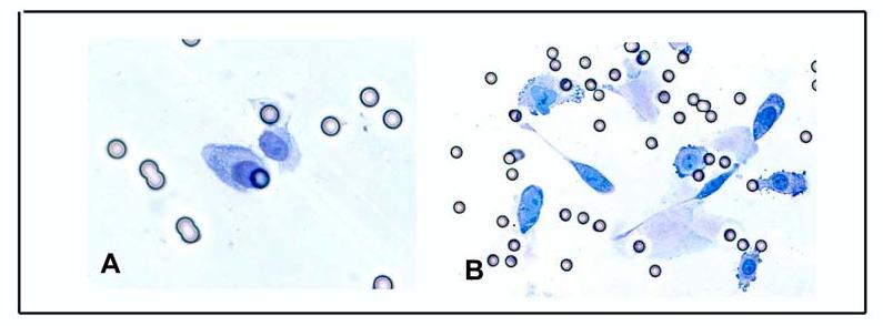
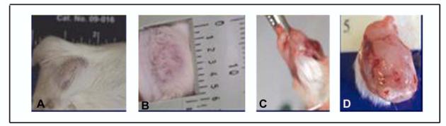
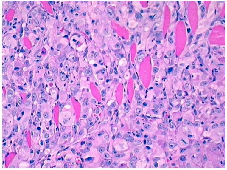
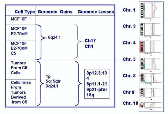
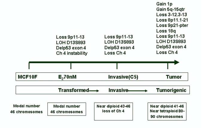
Estrogens are considered to play a major role in promoting the proliferation of both the normal and the neoplastic breast epithelium. Their role as breast carcinogens has long been suspected and recently confirmed by epidemiological studies. Three major mechanisms are postulated to be involved in their carcinogenic effects: stimulation of cellular proliferation through their receptor-mediated hormonal activity, direct genotoxic effects by increasing mutation rates through a cytochrome P450-mediated metabolic activation, and induction of aneuploidy. Recently it has been fully demonstrated that estrogens are carcinogenic in the human breast by testing in an experimental system the natural estrogen 17beta-estradiol (E(2)) by itself or its metabolites 2-hydroxy, 4-hydroxy, and 16-a-hydroxy-estradiol (2-OH-E(2), 4-OH-E(2), and 16-alpha-OH E(2)), respectively, by inducing neoplastic transformation of human breast epithelial cells (HBEC) MCF-10F in vitro to a degree at least similar to that induced by the chemical carcinogen benz(a)pyrene (BP). Neither Tamoxyfen (TAM) nor ICI-182,780 abrogated the transforming efficiency of estrogen or its metabolites. The E(2) induced expression of anchorage independent growth, loss of ductulogenesis in collagen, invasiveness in Matrigel, is associated with the loss of 9p11-13 and only invasive cells that exhibited a 4p15.3-16 deletion were tumorigenic. Tumors were poorly differentiated ER-alpha and progesterone receptor negative adenocarcinomas that expressed keratins, EMA and E-cadherin. The E(2) induced tumors and tumor-derived cell lines exhibited loss of chromosome 4, deletions in chromosomes 3p12.3-13, 8p11.1-21, 9p21-qter, and 18q, and gains in 1p, and 5q15-qter. The induction of complete transformation of the human breast epithelial cell MCF-10F in vitro confirms the carcinogenicity of E(2), supporting the concept that this hormone could act as an initiator of breast cancer in women. This model provides a unique system for understanding the genomic changes that intervene for leading normal cells to tumorigenesis and for testing the functional role of specific genomic events taking place during neoplastic transformation.
Figures
References
-
- Boyd S. An oophorectomy in cancer of the breast. Br. Med. J. 1900;2:1161–1167.
-
- Block G. Estrogen excretion following operative and irradiation castration in cases of mammary cancer. Surgery. 1958;43:415–422. - PubMed
-
- Green S, Walter P, Greene G, Krust A, Goffin C, Jensen E, Scrace G, Waterfield M, Chambon P. Cloning of the human estrogen receptor cDNA. J. Steroid Biochem. 1986;24:77–83. - PubMed
Publication types
MeSH terms
Substances
Grants and funding
LinkOut - more resources
Full Text Sources
Medical
Research Materials
