. 2006 Nov 17;24(4):581-92.
doi: 10.1016/j.molcel.2006.09.016.

SeqA blocking of DnaA-oriC interactions ensures staged assembly of the E. coli pre-RC

Affiliations

SeqA blocking of DnaA-oriC interactions ensures staged assembly of the E. coli pre-RC

Christian Nievera et al. Mol Cell. .

Abstract

DnaA occupies only the three highest-affinity binding sites in E. coli oriC throughout most of the cell cycle. Immediately prior to initiation of chromosome replication, DnaA interacts with additional recognition sites, resulting in localized DNA-strand separation. These two DnaA-oriC complexes formed during the cell cycle are functionally and temporally analogous to yeast ORC and pre-RC. After initiation, SeqA binds to hemimethylated oriC, sequestering oriC while levels of active DnaA are reduced, preventing reinitiation. In this paper, we investigate how resetting of oriC to the ORC-like complex is coordinated with SeqA-mediated sequestration. We report that oriC resets to ORC during sequestration. This was possible because SeqA blocked DnaA binding to hemimethylated oriC only at low-affinity recognition sites associated with GATC but did not interfere with occupation of higher-affinity sites. Thus, during the sequestration period, SeqA repressed pre-RC assembly while ensuring resetting of E. coli ORC.


Figures
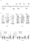
Figure 1
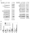
Figure 1. DnaA binding to oriC
A. Map of oriC with position and sequences of DnaA binding sites and GATC sequences indicated. GATCs associated with DnaA binding sites are underlined. Up and down arrows in the DnaA R- box and I-site recognition sequences indicate enhanced or reduced methylation by DMS. GATCs in three DnaA-ATP boxes (S-M sites) in the DNA Unwinding Region (DUE) are also marked. B. DMS modification patterns of oriC in vitro (lanes 1–9), after incubating supercoiled pAL40 minichromosme (200 fmole) with indicated concentrations of DnaA, or in vivo (lanes 10–18), on chromosomal oriC in exponentially growing cells or cells aligned at the time of initiation just prior to helicase loading, measured by LMPCR. Different primers and gel run times were used to resolve DnaA binding sites in three regions of oriC. DnaA binding sites and guanines at position 2 and 4 in the recognition site are marked. The Left, Middle, and Right 13 mer AT-rich sequences in the DUE are also labeled (L,M,R). The closed circle in the center panel indicates a putative, uncharacterized DnaA binding site, and the star in the third panel marks a G whose intensity changes slightly upon R3 binding. C. Relative intensities of DMS modification at G2 and G4 within 7 DnaA-binding sites. I1 and I3 sites lack guanine in position 2.
Figure 2
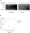
Figure 2. Binding of DnaA to oriC in vivo during the sequestration period and to hemimthylated supercoiled and linear oriC in vitro
A. Modification patterns following DMS treatment of chromosomal oriC (lanes 1,3) or oriC on pAL49 in dnaC(ts) cells 6 minutes after initiation (lanes 2, 4). Relative intensities of DMS-modified guanines in position 2 or 4 in the DnaA binding sites were quantified from scans of footprinting gels. B. pAL49 DNA was isolated from aligned dnaC(ts) cells at 2 minute intervals after a burst in synchronous initiation induced by return to permissive temperature, digested with Hph1, and analyzed using a Southern blot hybridized to an oriC probe. The percentage of cut and uncut pAL49 was quantified; 50% cutting indicates that 100% of the DNA was hemimethylated. The right panel shows the cutting pattern of fully methylated and hemimethylated oriC fragments generated by PCR as described in Experimental Procedures. C. Modification patterns following DMS treatment of purified supercoiled hemimethylated pAL49 (200 fmoles) incubated with 0 or 100 nM DnaA (lanes 1, 2, 6, 7), or hemimethylated PCR oriC fragment (200 fmoles) incubated with 0, 20, or 100 nM DnaA (lanes 3–5, 8–10). Positions of DnaA binding sites are shown. Relative intensities of DMS-modified guanines in position 2 or 4 in the DnaA binding sites in the presence and absence of DnaA were quantified from scans of footprinting gels.
Figure 3
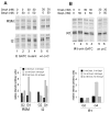
Figure 3. DnaA binds to lower affinity recognition sites shortly after initiation in cells lacking functional SeqA
A. Modification patterns following DMS treatment of purified chromosomal oriC (lanes 1,4) or oriC in dnaC(ts) seqA cells growing with multifork replication in rich media (minimal salts supplemented with glucose plus casamino acids), exponentially (lanes 2, 5) or 6 minutes after initiation was induced (lanes 3, 6). Relative intensities of DMS-modified guanines in position 2 or 4 in the DnaA binding sites were quantified from scans of footprinting gels. B. Modification patterns following DMS treatment of oriC in dnaC(ts) cells (lanes 1,3) or dnaC(ts) seqA cells (lanes 2,4) growing slowly in minimal salts supplemented with glycerol 6 minutes after initiation was induced. Relative intensities of DMS-modified guanines in position 2 or 4 in the DnaA binding sites were quantified from scans of footprinting gels.
Figure 4
Figure 4. Purified SeqA blocks DnaA from binding to lower affinity recognition sites on hemimethylated oriC
A. Modification patterns following DMS treatment of hemimethylated or fully methylated PCR oriC fragment (125 fmoles) incubated with indicated concentrations of SeqA and DnaA. When both proteins were present, SeqA was added first. Positions of DnaA binding sites are shown. (B) Relative intensities of DMS-modified guanines in position 2 or 4 in the DnaA binding sites after incubation with SeqA and DnaA were quantified from scans of footprinting gels.
Figure 5
Figure 5. SeqA inhibits DnaA-induced DNA strand separation of hemimethylated supercoiled DNA
A. Purified supercoiled hemimethylated pAL49 DNA (50 ng) was incubated with the indicated concentrations of SeqA and DnaA, and treated with P1 nuclease. 10 ng aliquots of the reaction were electrophoresed, and gels were stained with ethidium bromide. The positions of uncut supercoiled, nicked and linearized forms are shown. When both proteins are present, SeqA was added first. B. The amount of DNA in the linear band was quantified from the gels shown in (A).
Figure 6
Figure 6. SeqA blocks DnaA binding only from sites associated with a GATC
A. Modification patterns following DMS treatment of hemimethylated wild type oriC PCR fragment (right panel) or mutated oriC in which I2 no longer contained a GATC sequence (left). B. Modification patterns after DMS treatment of hemimethylated wild type (right panel) or mutated oriC in which a GATC sequence was put into R1 (left). Samples were incubated with indicated concentrations of SeqA and DnaA prior to DMS treatment. When both proteins were present, SeqA was added first. Positions of DnaA binding sites are shown. The lower panels show graphs of relative intensities of DMS-modified guanines in position 2 or 4 in the DnaA binding sites after incubation with SeqA and DnaA.

References

    1. Angers M, Cloutier JF, Castonguay A, Drouin R. Optimal conditions to use Pfu exo- DNA polymerase for highly efficient ligation-mediated polymerase chain reaction protocols. Nucl Acids Res. 2001;29:e83. - PMC - PubMed
    1. Bach T, Skarstad K. Re-replication from non-sequesterable origins generates three-nucleoid cells which divide asymmetrically. Mol Microbiol. 2004;51:1589–1600. - PubMed
    1. Baker TA, Bell SP. Polymerases and the replisome: machines within machines. Cell. 1998;92:295–305. - PubMed
    1. Bogan JA, Helmstetter CE. DNA sequestration and transcription in the oriC region of Escherichia coli. Mol Microbiol. 1997;26:889–896. - PubMed
    1. Boye E, Lobner-Olesen A, Skarstad K. Limiting DNA replication to once and only once. EMBO Rep. 2000;1:479–483. - PMC - PubMed

Publication types

MeSH terms

LinkOut - more resources