

PET/CT: will it change the way that we use CT in cancer imaging?
- PMID: 17114079
- PMCID: PMC1805068
- DOI: 10.1102/1470-7330.2006.9012
PET/CT: will it change the way that we use CT in cancer imaging?
Abstract
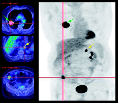
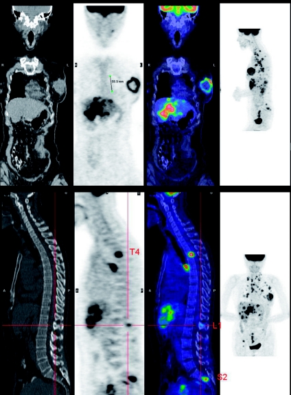
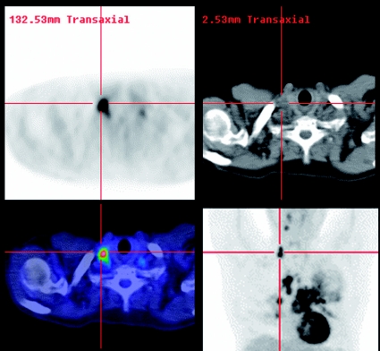
Accurate staging of cancer is of fundamental importance to treatment selection and planning. Current staging paradigms focus, first, on a detailed delineation of the primary tumour in order to determine its suitability for resection, and, thereafter, on assessment of the presence of metastatic spread that would alter the surgical approach, or mandate non-surgical therapies. This approach has, at its core, the assumption that the best, and sometimes the only, way to cure a patient of cancer is by surgical resection. Unfortunately, all non-invasive techniques in current use have imperfect ability to identify those primary tumours that are able to be completely excised, and even worse ability to define the extent of metastatic spread. Nevertheless, because of relatively low cost and widespread availability, computed tomography (CT) scanning is the preferred methodology for tumour, nodal and systemic metastasis (TNM) staging. This is often supplemented by other tests that have improved performance in particular staging domains. For example, magnetic resonance imaging (MRI), mammography, or endoscopic ultrasound may be used as complementary tests for T-staging; surgical nodal sampling for N-staging; and bone scanning, MRI or ultrasound for M-staging. Accordingly, many patients undergo a battery of investigations but, even then, are found to have been incorrectly staged based on subsequent outcomes. Even for those staged surgically, pathology can only identify metastases within the resection specimens and has no capability for detecting remote disease. As a result of this, many patients undergo futile operations for disease that could never have been cured by surgery. In the case of restaging, the situation is even worse. The sequelae of prior treatment can be difficult to differentiate from residual cancer and the likelihood of successful salvage therapy is even less than at presentation. More deleteriously, patients may be subjected to additional morbid treatments when cure has already been achieved. Thus, in post-treatment follow-up, the presence and extent of disease is equally critical to treatment selection and patient outcome as it is in primary staging. One of the major strengths of positron emission tomography (PET)/CT as a cancer staging modality is its ability to identify systemic metastases. At any phase of cancer evaluation, demonstration of systemic metastasis has profound therapeutic and prognostic implications. Only in the absence of systemic metastasis does nodal status become important, and only when unresectable nodal metastasis has been excluded does T-stage become important. There are now accumulating data that PET/CT could be used as the first, rather than the last test to assess M- and N-stage for evaluating cancers with an intermediate to high pre-test likelihood of metastatic disease based on poor long-term survival. In this scenario, there is great opportunity for subsequently selecting and tailoring the performance of anatomically based imaging modalities to define the structural relations of abnormalities identified by PET, when this information would be of relevance to management planning. Primary staging of oesophageal cancer and restaging of colorectal cancer are illustrative examples of a new paradigm for cancer imaging.
(c) International Cancer Imaging Society.
Figures
References
-
- Jones LA, Chilton JA, Hajek RA, Iammarino NK, Laufman L. Between and within: international perspectives on cancer and health disparities. J Clin Oncol. 2006;24:2204–8. - PubMed
-
- Bosanquet N, Sikora K. The economics of cancer care in the UK. Lancet Oncol. 2004;5:568–74. - PubMed
-
- Edwards BK, Brown ML, Wingo PA, et al. Annual report to the nation on the status of cancer, 1975–2002, featuring population-based trends in cancer treatment. J Natl Cancer Inst. 2005;97:1407–27. - PubMed
-
- Gambhir SS, Czernin J, Schwimmer J, Silverman DH, Coleman RE, Phelps ME. A tabulated summary of the FDG PET literature. J Nucl Med. 2001;42:1S–93S. - PubMed
-
- Beyer T, Townsend DW, Brun T, et al. A combined PET/CT scanner for clinical oncology. J Nucl Med. 2000;41:1369–79. - PubMed
MeSH terms
Substances
LinkOut - more resources
Full Text Sources
Medical
Research Materials