. 2007 Feb 15;92(4):1409-18.
doi: 10.1529/biophysj.106.087197. Epub 2006 Nov 17.

The cooperative response of synaptotagmin I C2A. A hypothesis for a Ca2+-driven molecular hammer

Affiliations

The cooperative response of synaptotagmin I C2A. A hypothesis for a Ca2+-driven molecular hammer

Jill A Kertz et al. Biophys J. .

Abstract

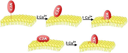
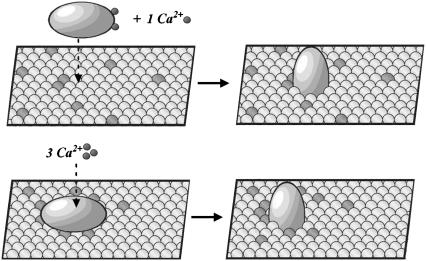
In the current understanding of exocytosis at the nerve terminal, the C2 domain of synaptotagmin (C2A) is presumed to bind Ca2+ and the membrane in a stepwise fashion: cation then membrane as cation increases the affinity of protein for membrane. Fluorescence spectroscopy data were gathered over a variety of lipid and Ca2+ concentrations, enabling the rigorous application of microscopic binding models derived from partition functions to differentiate between Ca2+ and phosphatidylserine contributions to binding. The data presented here are in variance with previously published models, which were based on the Hill approximation. Rather, the data are consistent with two forms of cooperativity that modulate the responsiveness of C2A: in Ca2+ binding to a network of three cation sites and in interaction with the membrane surface. We suggest synaptotagmin I C2A is preassociated with the synaptic vesicle membrane or nerve terminal. In this state, upon Ca2+ influx the protein will bind the three Ca2+ ions immediately and with high cooperativity. Thus, membrane association creates a high-affinity Ca2+ switch that is the basis for the role of synaptotagmin I in Ca2+-regulated exocytosis. Based on this model, we discuss the implications of protein-induced phosphatidylserine demixing to the exocytotic process.


Figures

FIGURE 1
FIGURE 1
(top) ‘Solution-first’ model. (bottom) Proposed model.
FIGURE 2
FIGURE 2
(A) Binding of Tb3+ to synaptotagmin C2A in the absence of lipid. The signal arising from quenching the intrinsic protein fluorescence was used. The loss of fluorescence was normalized and expressed as fraction bound. The added total ligand was corrected to free ligand, taking into account the total number of ligands occupying the protein at saturation and the concentration of protein (0.2 μM). Data were corrected for two cation-binding sites. All titrations were carried out in decalcified 2 mM MOPS, 100 mM KCl, pH 7.5. The line is Eq. 1 applied to the partition function q0. (B) Binding of Tb3+ to synaptotagmin C2A (0.55 μM) in the presence of 40 μM total POPC/POPS (16:0,18:1PC:16:0,18:1PS) (75:25). This corresponds to 20 μM POPC/POPS available for binding on the outer leaflet of the large unilamellar vesicles. The fitted line is Eq. 1 applied to the global partition function Q. K and σ are held constant from Fig. 2 A and KL = 0.67 μM−1.
FIGURE 3
FIGURE 3
Binding of synaptotagmin C2A to POPC/POPS (75:25) or POPC/POPS/dansylPE (65:25:10) in the presence of subsaturating Tb3+ (9.1 μM, A) or Ca2+ (2 μM, B). The lipid titrant was corrected for free lipid concentration assuming membrane coverage by C2A corresponds to five lipids and for only outer leaflet lipid available for binding. The same partition function and constants were used to fit both binding isotherms with the corresponding constant cation concentration, 9.1 μM or 2 μM, for Fig. 3, A or B. Protein concentration was 0.55 μM for (A) and 0.2 μM for (B).
FIGURE 4
FIGURE 4
(A) Dependence of the lipid-binding constant of synaptotagmin I C2A on the percentage of PS in a binary mixture of POPC/POPS. KL is presented as the logarithm of the dissociation constant (1/KL). The logarithm is provided as base 10 for ease of comparison of the values to other figures. Protein concentrations were varied from 0.2–0.55 μM. (B) Binding of synaptotagmin C2A to POPC/POPS/dansylPE (65:25:10). The lipid-binding constants were calculated through application of Eq. 1 to partition functions. The global partition function Q from setting [Tb3+] = 0 in Eq. 3, then reverts to 1 + KL(L). The lipid titrant was corrected for free lipid concentration as above, accounting for protein concentration, outer leaflet lipids, and membrane coverage by C2A of five lipids.
FIGURE 5
FIGURE 5
Binding of Tb3+ to 0.4 μM synaptotagmin C2A in the presence of 400 μM total POPC (A) and of POPC to C2A in the presence of 9.1 μM Tb3+ (B). Data were corrected for as in Figs. 2 and 3.
FIGURE 6
FIGURE 6
(top) ‘Solution-first’ model which does not involve PS-demixing upon C2A binding to membrane. (bottom) Proposed model incorporating PS demixing. PS is shown in green and the ‘bulk’ membrane, which in our model system is represented by PC, is shown in yellow.
FIGURE A1
FIGURE A1
Binding of Tb3+ to synaptotagmin C2A in the presence of 40 μM total POPC/POPS (16:0,18:1PC:16:0,18:1PS) (75:25). Details are the same as Fig. 2 A. The lines correspond to Eq. 1 applied to the partition function (Qsolution-first = 1 + 2K[Tb3+] + σK2[Tb3+]2 + KL[L](σK2[Tb3+]2 + σρK3[Tb3+]3) (Eq. A4). K and σ are held constant from Fig. 2. The solid line corresponds to the use of the KL also ascertained from the fit of the lipid titration data of Fig. 2 A. The dashed line corresponds to fitting the data with a ρ = 19,000 (from the membrane titration), and the solid line corresponded with a ρ = 168.

References

    1. Fernandez-Chacon, R., A. Königstorfer, S. H. Gerber, J. Garcia, M. F. Matos, C. F. Stevens, N. Brose, J. Rizo, C. Rosenmund, and T. C. Sudhof. 2001. Synaptotagmin I functions as a calcium regulator of release probability. Nature. 410:41–49. - PubMed
    1. Fernandez-Chacon, R., O.-H. Shin, A. Königstorfer, M. F. Matos, A. C. Meyer, J. Garcia, S. H. Gerber, J. Rizo, T. C. Sudhof, and C. Rosenmund. 2002. Structure/function analysis of Ca2+ binding to the C2A domain of synaptotagmin 1. J. Neurosci. 22:8438–8446. - PMC - PubMed
    1. Sorensen, J. B., R. Fernandez-Chacon, T. C. Sudhof, and E. Neher. 2003. Examining synaptotagmin 1 function in dense core vesicle exocytosis under direct control of Ca2+. J. Gen. Physiol. 122:265–270. - PMC - PubMed
    1. Shin, O.-H., J.-S. Rhee, J. Tang, S. Sugita, C. Rosenmund, and T. C. Sudhof. 2003. Sr2+ binding to the Ca2+ binding site of the synaptotagmin 1 C2B domain triggers fast exocytosis without stimulating SNARE interactions. Neuron. 37:99–109. - PubMed
    1. Sudhof, T. C. 2004. The synaptic vesicle cycle. Annu. Rev. Neurosci. 27:509–547. - PubMed

Publication types

MeSH terms

LinkOut - more resources