. 2007 Feb;189(3):902-10.
doi: 10.1128/JB.01513-06. Epub 2006 Nov 17.

Physical and functional interactions between Escherichia coli MutY glycosylase and mismatch repair protein MutS

Affiliations

Physical and functional interactions between Escherichia coli MutY glycosylase and mismatch repair protein MutS

Haibo Bai et al. J Bacteriol. 2007 Feb.

Abstract

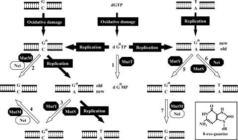
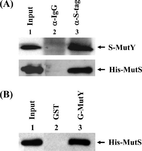
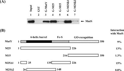
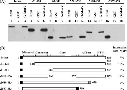
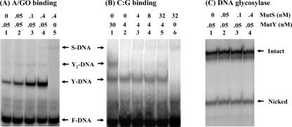
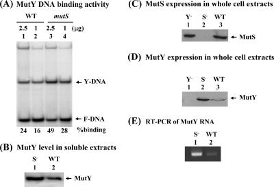
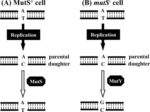
Escherichia coli MutY and MutS increase replication fidelity by removing adenines that were misincorporated opposite 7,8-dihydro-8-oxo-deoxyguanines (8-oxoG), G, or C. MutY DNA glycosylase removes adenines from these mismatches through a short-patch base excision repair pathway and thus prevents G:C-to-T:A and A:T-to-G:C mutations. MutS binds to the mismatches and initiates the long-patch mismatch repair on daughter DNA strands. We have previously reported that the human MutY homolog (hMYH) physically and functionally interacts with the human MutS homolog, hMutSalpha (Y. Gu et al., J. Biol. Chem. 277:11135-11142, 2002). Here, we show that a similar relationship between MutY and MutS exists in E. coli. The interaction of MutY and MutS involves the Fe-S domain of MutY and the ATPase domain of MutS. MutS, in eightfold molar excess over MutY, can enhance the binding activity of MutY with an A/8-oxoG mismatch by eightfold. The MutY expression level and activity in mutS mutant strains are sixfold and twofold greater, respectively, than those for the wild-type cells. The frequency of A:T-to-G:C mutations is reduced by two- to threefold in a mutS mutY mutant compared to a mutS mutant. Our results suggest that MutY base excision repair and mismatch repair defend against the mutagenic effect of 8-oxoG lesions in a cooperative manner.


Figures

FIG. 1.
FIG. 1.
8-OxoG repair in E. coli. MutT, MutM, MutS, MutY, and Nei (endonuclease VIII) are involved in defending against the mutagenic effects of 8-oxoG lesions (structure is shown in the inset). The MutT protein hydrolyzes 8-oxo-dGTP (dGoTP) to 8-oxo-dGMP (dGoMP) and pyrophosphate (reaction 1). GO (Go) in DNA can be derived from oxidation of guanine or misincorporation of dGoTP during replication. The MutM glycosylase removes GO adducts while it is paired with cytosine (reactions 2, 4, and 7). Nei can function as a backup for MutM to remove GO from GO/C. When C/GO is not repaired by MutM, adenines are frequently incorporated opposite GO bases by DNA polymerase III during DNA replication. A/GO mismatches are repaired to C/GO by the MutY-dependent or MutS-dependent pathway (reaction 3). When dGoTP is incorporated opposite adenine during DNA replication, MutY repair on GO/A can cause more mutation (reaction 5) while GO/A repair by MutS and Nei can reduce mutation (reaction 6). This figure is adapted from the work of Lu et al. with permission of the publisher (41).
FIG. 2.
FIG. 2.
Physical interaction of MutY and MutS. (A) Coimmunoprecipitation of MutY with MutS. Immunoprecipitation was performed with rabbit anti-mouse (lane 2) or S-tag (lane 3) antibody and extracts from BL21(DE3) cells expressing S-tagged MutY and His-tagged MutS proteins. Western blotting was performed with S-tag antibody (upper panel) or with His-tag antibody (lower panel). Lane 1 of the upper panel represents 15% of input extract, and lane 1 of the lower panel represents 0.5% of input extract. (B) Binding of MutS to GST-tagged MutY. GST (lane 2) or GST-MutY (lane 3) immobilized to glutathione-Sepharose beads was used to pull down His-tagged MutS protein. The pellets were fractionated on an 8% SDS-polyacrylamide gel followed by Western blot analysis to detect MutS with the antibody to His tag. Lane 1 contains 10% of input MutS protein.
FIG. 3.
FIG. 3.
Determination of regions of MutY involved in MutS binding. (A) Various GST-MutY constructs were immobilized to glutathione-Sepharose beads and used to pull down His-tagged MutS protein. A control was run concurrently with immobilized GST alone (lane 2). Lane 1 contains 10% of input His-tagged MutS protein. The same amounts of GST fusion proteins were used in the experiments by normalizing with the corresponding protein bands on a Coomassie blue-stained 12% SDS-polyacrylamide gel (data not shown). The pellets were fractionated on an 8% SDS-polyacrylamide gel followed by Western blot analysis with the antibody to His tag. (B) Graphic depiction of GST-MutY constructs and the binding with MutS proteins. The region shaded in black is the six-helix barrel (residues 26 to 134). The Fe-S domain consists of residues 1 to 25 and 135 to 226. The C-terminal domain of MutY is involved in GO recognition. The portions of protein present in the MutY deletion constructs are indicted by boxes and numbers of amino acid residues. The strength of binding, presented at the right, was calculated as the ratio of the amount of MutS in the pellet to that of input MutS (10% in lane 1).
FIG. 4.
FIG. 4.
Determination of regions within MutS involved in MutY binding. (A) The GST pull-down assays were performed with different MutS constructs as indicated and GST-MutY immobilized on glutathione-Sepharose (lanes 3, 6, 9, 12, 15, and 18). Controls were run concurrently with immobilized GST alone for each MutS construct (lanes 2, 5, 8, 11, 14, and 17). Ten percent of each input MutS construct was loaded to lanes 1, 4, 7, 10, 13, and 16. Western blot analyses of the pellets were performed with antibody to His tag. (B) Graphic depiction of MutS constructs and the binding to GST-MutY fusion protein. The amino-terminal mismatch recognition domain (residues 2 to 115), the connector domain (residues 116 to 266), the core domain (residues 267 to 443 and 504 to 567), the ATPase domain (residues 568 to 765), and the HTH domain (residues 766 to 800) are indicated. Residues 444 to 503 are the clamp domain. The names of MutS domains with their corresponding residues were adapted from the work of Lamers et al. (30). In addition, the extreme C-terminal domain (residues 801 to 853) is responsible for the tetramer formation (5). The portions of protein present in the MutS deletion constructs are indicted by boxes and numbers of amino acid residues. The black and gray boxes at the N or C terminus of MutS represent His tag and an additional 13 amino acids, respectively. The strength of binding, presented at the right, was calculated as the ratio of the amount of MutS in the pellet to that of input MutS constructs.
FIG. 5.
FIG. 5.
MutS stimulates MutY binding activity towards A/8-oxoG-containing DNA. (A) DNA substrates (0.09 nM) containing A/GO mismatches were incubated with MutY (0.05 nM) (lane 1) and increasing amounts of MutS (lanes 2 to 4). The reaction mixture in lane 5 contains 0.4 nM MutS and no MutY. The samples were fractionated on a nondenaturing 4% polyacrylamide gel. The arrows indicate the positions of MutY-DNA complex (Y-DNA), MutS-DNA complex (S-DNA), and free DNA substrate (F-DNA). The faint band (marked by Y2-DNA) between the Y-DNA and S-DNA bands may be a complex of MutY dimer and DNA (31). (B) Homoduplex DNA substrates (0.09 nM) containing C:G were incubated with MutY as indicated (lanes 1 and 2) and increasing amounts of MutS (lanes 3 to 5). The reaction mixture in lane 6 contains 32 nM MutS and no MutY. Reaction mixtures are similar to those of panel A. (C) A/8-oxoG-containing DNA substrates (0.09 nM) were incubated with MutY (0.05 nM) (lane 1) and increasing amounts of MutS (lanes 2 to 4). After MutY incubation, the reaction mixtures were heated at 90°C for 30 min in the presence of 0.1 M NaOH. The samples were fractionated on a denaturing 14% polyacrylamide gel. The arrows indicate the positions of intact and nicked DNA.
FIG. 6.
FIG. 6.
The DNA binding activity and expression level of MutY are upregulated in the mutS cells. (A) MutY binding activity towards A/8-oxoG-containing DNA with extracts from AB1157 (wild type [WT], lanes 1 and 2) and KM75 (mutS, lanes 3 and 4). The assay was performed with 1 and 2.5 μg of cell extracts. The samples were fractionated on nondenaturing 6% polyacrylamide gels. The arrows indicate the positions of MutY-DNA complex (Y-DNA) and free DNA substrate (F-DNA). The numbers at the bottom of the figure indicate the percentages of DNA substrate bound by MutY. (B) Western blot analysis for MutY with 80 μg soluble extracts from AB1157 (WT) and KM75 (mutS, S) cells. (C) Western blot analysis for MutS was performed with 60 μg total cell proteins from AB1157 (WT), KM75 (mutS, S), and GM7724 (mutY, Y) cells. (D) Western blot analysis for MutY was performed with 60 μg total cell proteins from AB1157 (WT), KM75 (mutS, S), and GM7724 (mutY, Y) cells. (E) MutY mRNA was quantitated by reverse transcriptase PCR (RT-PCR) with isolated RNA from AB1157 (WT) and KM75 (mutS, S) cells. The DNA products were resolved on a 1% agarose gel and visualized by staining with ethidium bromide.
FIG. 7.
FIG. 7.
Overproduction of MutY in E. coli mutS cells may have a mutagenic effect on A:T-to-G:C transitions. (A) In MutS+ cells, A/C mismatches generated during DNA replication are mainly repaired by MutS-dependent mismatch repair. (B) In mutS cells, overproduced MutY can remove A from an A/C mismatch in which A is on the parental strand and thus cause an A:T-to-G:C transition.

References

    1. Al-Tassan, N., N. H. Chmiel, J. Maynard, N. Fleming, A. L. Livingston, G. T. Williams, A. K. Hodges, D. R. Davies, S. S. David, J. R. Sampson, and J. P. Cheadle. 2002. Inherited variants of MYH associated with somatic G:C to T:A mutations in colorectal tumors. Nat. Genet. 30:227-232. - PubMed
    1. Au, K. G., M. Cabrera, J. H. Miller, and P. Modrich. 1988. Escherichia coli mutY gene product is required for specific A/G to C:G mismatch correction. Proc. Natl. Acad. Sci. USA 85:9163-9166. - PMC - PubMed
    1. Au, K. G., S. Clark, J. H. Miller, and P. Modrich. 1989. Escherichia coli mutY gene encodes an adenine glycosylase active on G/A mispairs. Proc. Natl. Acad. Sci. USA 86:8877-8881. - PMC - PubMed
    1. Biswas, I., G. Obmolova, M. Takahashi, A. Herr, M. A. Newman, W. Yang, and P. Hsieh. 2001. Disruption of the helix-U-turn-helix motif of MutS protein: loss of subunit dimerization, mismatch binding and ATP hydrolysis. J. Mol. Biol. 305:805-816. - PubMed
    1. Bjornson, K. P., L. J. Blackwell, H. Sage, C. Baitinger, D. Allen, and P. Modrich. 2003. Assembly and molecular activities of the MutS tetramer. J. Biol. Chem. 278:34667-34673. - PubMed

Publication types

MeSH terms

LinkOut - more resources