. 2006 Nov 28;103(48):18154-9.
doi: 10.1073/pnas.0602235103. Epub 2006 Nov 17.

Prolyl hydroxylase-1 negatively regulates IkappaB kinase-beta, giving insight into hypoxia-induced NFkappaB activity

Affiliations

Prolyl hydroxylase-1 negatively regulates IkappaB kinase-beta, giving insight into hypoxia-induced NFkappaB activity

Eoin P Cummins et al. Proc Natl Acad Sci U S A. .

Abstract

Hypoxia is a feature of the microenvironment of a growing tumor. The transcription factor NFkappaB is activated in hypoxia, an event that has significant implications for tumor progression. Here, we demonstrate that hypoxia activates NFkappaB through a pathway involving activation of IkappaB kinase-beta (IKKbeta) leading to phosphorylation-dependent degradation of IkappaBalpha and liberation of NFkappaB. Furthermore, through increasing the pool and/or activation potential of IKKbeta, hypoxia amplifies cellular sensitivity to stimulation with TNFalpha. Within its activation loop, IKKbeta contains an evolutionarily conserved LxxLAP consensus motif for hydroxylation by prolyl hydroxylases (PHDs). Mimicking hypoxia by treatment of cells with siRNA against PHD-1 or PHD-2 or the pan-prolyl hydroxylase inhibitor DMOG results in NFkappaB activation. Conversely, overexpression of PHD-1 decreases cytokine-stimulated NFkappaB reporter activity, further suggesting a repressive role for PHD-1 in controlling the activity of NFkappaB. Hypoxia increases both the expression and activity of IKKbeta, and site-directed mutagenesis of the proline residue (P191A) of the putative IKKbeta hydroxylation site results in a loss of hypoxic inducibility. Thus, we hypothesize that hypoxia releases repression of NFkappaB activity through decreased PHD-dependent hydroxylation of IKKbeta, an event that may contribute to tumor development and progression through amplification of tumorigenic signaling pathways.


Conflict of interest statement

The authors declare no conflict of interest.

Figures

Fig. 1.
Fig. 1.
Hypoxia activates NFκB. (A) HeLa cells transfected with an NFκB-luciferase reporter and exposed to 1% O2 (0–48 h) demonstrate increased NFκB activity. (B) Nuclear extracts from HeLa cells exposed to preconditioned hypoxic medium (1% O2; 4 h) demonstrate increased NFκB DNA binding. (C) HeLa cells exposed to graded hypoxia (21–1% O2; 24 h) demonstrate decreased extracellular pO2 values as measured by fluorescence quenching oxymetry and increased NFκB and HIF-1α DNA binding activity (D and E).
Fig. 2.
Fig. 2.
NFκB DNA-binding assay in HeLa cells exposed to preconditioned hypoxic medium (1% O2) for 1 h before TNFα treatment (0.01–0.1 ng/ml for an additional hour) demonstrates enhanced NFκB-nuclear binding when compared with normoxic controls.
Fig. 3.
Fig. 3.
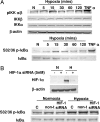
Hypoxia activates NFκB through the IKK complex. (A) HeLa cells exposed to preconditioned normoxic (N; 21% O2) or hypoxic (H; 1% O2) media (5–120 min) demonstrate temporally sequential activation of IKKα/β, phosphorylation of IκBα, and degradation of IκBα. (B) HeLa cells transfected with siRNA against HIF-1α or nontarget siRNA exposed to hypoxia (1% O2; 24 h) demonstrate effective HIF-1α knockdown. siRNA against HIF-1α does not prevent hypoxia-induced phosphorylation of IκBα.
Fig. 4.
Fig. 4.
PHDs suppress NFκB activity. (A) IKKα and IKKβ but not NEMO (not shown) contain conserved LxxLAP motifs. (B) HeLa cells transiently transfected with PHD siRNA demonstrate effective isoform-specific knockdown by quantitative RT-PCR. Data are shown from a representative experiment (n = 3 in total). (C) HeLa cells were cotransfected with isoform-specific PHD siRNAs (20 nM), the reporter vector (pNFκB-LUC), and 100 ng of a β-galactosidase construct. As a control for NFκB activation, cells were incubated with TNFα (10 ng/ml). Forty-eight hours after transfection, cells were lysed and luciferase and β-galactosidase activity were measured (37). Results are expressed as the fold induction over control. (D) HeLa cells were transiently cotransfected with pEGLN2-FLAG (PHD1; 0.15–0.3 μg) or empty pcDNA (0.15–0.3 μg) vector and pNFκB-LUC. After transfection, cells were treated with TNFα (0.1 ng/ml) for 24 h. Whole-cell lysates were prepared, and a luciferase assay was carried out. Results are protein-normalized RLU values expressed as fold over basal luciferase activity. (E) HeLa cells were transiently transfected with an NFκB-promoter reporter construct. After transfection, cells were exposed to DMSO-vehicle (control) or 1 mM DMOG and maintained at 21% O2 for 24 h. Results shown are protein normalized RLU values. (F) Immunoblotting for anti-phospho-S32/36 IκBα and total IκBα was carried out on DMOG-treated HeLa cells (1 mM; 2–75 min). (G) HeLa cells were exposed to DMOG (1 mM, 24 h) or vehicle. Whole-cell extracts were immunoblotted for HIF-1α, Cox-2, or β-actin. (H) Three PHD isoforms were knocked down in HeLa cells as described above, and Cox-2 expression was determined by Western blot analysis.
Fig. 5.
Fig. 5.
The cellular pool of IKKβ is increased in hypoxia. HeLa (A) and THP-1 and CaCo-2 (B) cells were exposed to graded hypoxia (21–1% O2) for 24 h or instantaneous hypoxia (1% O2) for 0–6 h. Whole-cell extracts were immunoblotted for IKKβ, NEMO, and β-actin where indicated. (C) HeLa cells were transiently transfected with 1 μg of wild-type IKKβ or P191A mutant IKKβ. Forty-eight hours after transfection, the cells were maintained in normoxia (N; 21% O2) or exposed to preconditioned hypoxic medium (H; 3% O2) for 2 h. Cytosolic extracts were immunoblotted for IKKβ and β-actin. Alterations in protein expression were measured semiquantitatively by using densitometric analysis (n = 4). (D) pVHL was immunoprecipitated from whole HeLa cell extracts (500 or 1,000 μg of total protein), and immunoprecipitates were immunoblotted for IKKβ and hexokinase (similarly sized negative control). (E) NETN lysates from HeLa cells transiently overexpressing PHD-1-FLAG were immunoprecipitated by using a specific anti-FLAG resin. Immunoprecipitates were immunoblotted for IKKβ, IKKα, FLAG, and hexokinase.

References

    1. Harris AL. Nat Rev Cancer. 2002;2:38–47. - PubMed
    1. Schofield CJ, Ratcliffe PJ. Nat Rev Mol Cell Biol. 2004;5:343–354. - PubMed
    1. Cummins EP, Taylor CT. Pflügers Arch. 2005;450:363–371. - PubMed
    1. Karin M, Greten FR. Nat Rev Immunol. 2005;5:749–759. - PubMed
    1. Berra E, Ginouves A, Pouyssegur J. EMBO Rep. 2006;7:41–45. - PMC - PubMed

Publication types

LinkOut - more resources