. 2006 Nov;18(11):3121-31.
doi: 10.1105/tpc.106.042671. Epub 2006 Nov 17.

Psb27, a cyanobacterial lipoprotein, is involved in the repair cycle of photosystem II

Affiliations

Psb27, a cyanobacterial lipoprotein, is involved in the repair cycle of photosystem II

Marc M Nowaczyk et al. Plant Cell. 2006 Nov.

Abstract

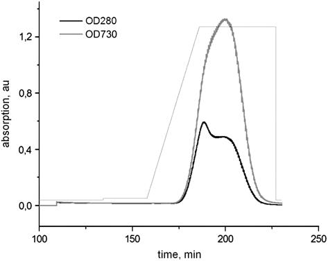
Photosystem II (PSII) performs one of the key reactions on our planet: the light-driven oxidation of water. This fundamental but very complex process requires PSII to act in a highly coordinated fashion. Despite detailed structural information on the fully assembled PSII complex, the dynamic aspects of formation, processing, turnover, and degradation of PSII with at least 19 subunits and various cofactors are still not fully understood. Transient complexes are especially difficult to characterize due to low abundance, potential heterogeneity, and instability. Here, we show that Psb27 is involved in the assembly of the water-splitting site of PSII and in the turnover of the complex. Psb27 is a bacterial lipoprotein with a specific lipid modification as shown by matrix-assisted laser-desorption ionization time of flight mass spectrometry. The combination of HPLC purification of four different PSII subcomplexes and (15)N pulse label experiments revealed that lipoprotein Psb27 is part of a preassembled PSII subcomplex that represents a distinct intermediate in the repair cycle of PSII.


Figures

Figure 1.
Figure 1.
Purification of His-Tagged PSII Complexes. His-tagged PSII complexes were eluted with a linear gradient of 1 to 100 mM histidine (light-gray line) from a Ni-NTA sepharose column. The elution profile is shown at 280 nm (arbitrary units [au]; black line) and at 730 nm (dark-gray line). For details, see Methods.
Figure 2.
Figure 2.
Purification and Characterization of His-Tagged PSII Complexes. (A) Elution profile from an UNO Q6 ion exchange column (Bio-Rad) of prepurified His-tagged PSII complexes. Individual PSII subfractions: 1, PSIIM(low); 2, PSIIM(high); 3, PSIID(high); 4, PSIID(low). (B) Monomer/dimer analysis of PSII subcomplexes by native-PAGE. (C) SDS-PAGE profiles showing the differences in subunit (SU) composition of the four IEC subfractions. Each lane was loaded with 2 μg Chl. For details, see Methods.
Figure 3.
Figure 3.
P680+QA - P680QA Difference Spectra of PSII Core Complexes from PSIIM(low), PSIIM(high), PSIID(high), and PSIID(low). The spectra have been recorded at 77K and are normalized to the bleaching minimum. While the main bleaching at 674 nm could be assigned to the oxidation of P680, the absorbance increase at 680 nm and the absorbance decrease at 684 nm have been assigned to an electrochromic blue shift of the QY absorption band of the accessory chlorophyll (Diner et al., 2001). Triangles, PSIIM(low); squares, PSIIM(high); circles, PSIID(high); inverse triangles, PSIID(low).
Figure 4.
Figure 4.
Processing of the D1 Protein. Intact PSIIM(low) (gray) and PSIID(high) (black) complexes were analyzed in the mass-to-charge (m/z) range between 37.000 and 41.000 by MALDI-TOF. The assignment of the peaks is based on the calculated masses for unprocessed D1 (SwissProt primary accession number P0A444; molecular mass 39,605 D), processed D1 (38,109 D), and D2 (Q8CM25; 39,230 D).
Figure 5.
Figure 5.
SDS-PAGE Analysis of PSII/Psb27 Complexes Reconstituted with the Extrinsic PSII Proteins PsbO, PsbV, and PsbU. Lane 1, PSIID(high); lane 2, PSIID(high) after treatment with 1 M CaCl2 to remove the extrinsic proteins PsbO, PsbV, and PsbU; lane 3, released extrinsic proteins; lane 4, PSIID(high) after treatment with 1 M CaCl2 and reconstitution with the released extrinsic proteins; lane 5, PSIIM(low)-Psb27 complex; lane 6, PSIIM(low)-Psb27 after reconstitution with PsbO, PsbV, and PsbU.
Figure 6.
Figure 6.
Signal Peptides of Psb27 from Different Species. Characteristic features of the signal peptides are labeled as follows: horizontal bar, region of hydrophobic and uncharged residues; boxed region, Lipobox motif with the invariant Cys residue (+1).
Figure 7.
Figure 7.
MS Analysis of Psb27. Intact PSIIM(low) complexes were analyzed by MALDI-TOF with or without Lipolase treatment to determine the accurate mass of Psb27 (SwissProt primary accession number Q8DG60) and to probe the prediction of the posttranslational lipid modification. Masses in a m/z range of 12,000 to 14,000 were detected before (native Psb27; black) and after 5 min (P1; grey) or 30 min (P2; light gray) of Lipolase treatment (for details, see Methods). The inset shows the model and the calculated (total) mass of the best-suited Psb27 lipid modification based on the masses determined at each stage.
Figure 8.
Figure 8.
Analysis of 15N Incorporation into the D1 Subunit of PSII. T. elongatus cells, grown in a medium with the natural nitrogen isotope 14N (99.6%), were exposed to a medium highly enriched in the stable isotope 15N (>98%) at the mid-log growth phase. Cells were harvested 3 and 10 h after the 15N pulse, followed by an immediate preparation of PSII subcomplexes. Subunits of each PSII subcomplex were separated by SDS-PAGE, and the D1 subunit was subjected to tryptic digestion and MALDI-TOF analysis (for details, see Methods). The top row (P1) shows the incorporation of 15N into a peptide (calculated mass 2188.3 D) from the D1 subunit (SwissProt primary accession number P0A444) of the PSIIM(low)-Psb27 complex 3 h (left column) and 10 h (right column) after the pulse. Spectra of the bottom row (P3) show the corresponding peptide from the D1 subunit of the PSIID(high) complex. The percentage of newly synthesized D1 protein was calculated from the spectra. A peak corresponding to an autocatalytic product of trypsin is marked by an asterisk.
Figure 9.
Figure 9.
Analysis of 15N Incorporation into Different Subunits of PSIIM(low). T. elongatus cells were harvested 24 h after the 15N pulse, followed by preparation of PSIIM(low) and SDS-PAGE analysis. Several subunits were subjected to tryptic digestion and analyzed by MALDI-TOF. The incorporation of 15N was followed into peptides of PsbE (SwissProt primary accession number Q8DIP0; molecular mass 1806.9 m/z), D1 (P0A444; 2188.3 m/z), D2 (Q8CM25; 1257.3 m/z), CP43 (Q8DIF8; 1681.7 m/z), and CP47 (Q8DIQ1; 1864.0 m/z), and the amount of newly synthesized protein was calculated.
Figure 10.
Figure 10.
Schematic Model for the Transient PSII Subcomplexes Isolated in This Study. Upon damage by light, the highly active dimeric PSII complex [PSIID(high)] is transformed into a subfraction of less-active PSII [PSIID(low)]. In the next step, the D1 subunit and others are exchanged before the inactive monomeric PSII-Psb27 complex [PSIIM(low)] could be isolated. This complex, which performs light-induced charge separation but is incapable of water splitting as it lacks subunits PsbO, PsbU, and PsbV, is turned into the active PSII monomer [PSIIM(high)] after release of Psb27 and assembly of the water-splitting site. Finally, two active monomers merge to a highly active dimeric PSII complex [PSIID(high)].

References

    1. Anbudurai, P.R., Mor, T.S., Ohad, I., Shestakov, S.V., and Pakrasi, H.B. (1994). The ctpA gene encodes the C-terminal processing protease for the D1 protein of the photosystem II reaction center complex. Proc. Natl. Acad. Sci. USA 91 8082–8086. - PMC - PubMed
    1. Anderson, J.M., Park, Y.I., and Chow, W.S. (1997). Photoinactivation and photoprotection of photosystem II in nature. Physiol. Plant 100 214–223.
    1. Aro, E.M., Suorsa, M., Rokka, A., Allahverdiyeva, Y., Paakkarinen, V., Saleem, A., Battchikova, N., and Rintamaki, E. (2005). Dynamics of photosystem II: A proteomic approach to thylakoid protein complexes. J. Exp. Bot. 56 347–356. - PubMed
    1. Bendtsen, J.D., Nielsen, H., von Heijne, G., and Brunak, S. (2004). Improved prediction of signal peptides: SignalP 3.0. J. Mol. Biol. 340 783–795. - PubMed
    1. Chen, H., Zhang, D., Guo, J., Wu, H., Jin, M., Lu, Q., Lu, C., and Zhang, L. (2006). A Psb27 homologue in Arabidopsis thaliana is required for efficient repair of photodamaged photosystem II. Plant Mol. Biol. 61 567–575. - PubMed

Publication types

MeSH terms

LinkOut - more resources