. 2006 Dec 1;177(11):7802-10.
doi: 10.4049/jimmunol.177.11.7802.

Generation and growth of CD28nullCD8+ memory T cells mediated by IL-15 and its induced cytokines

Affiliations

Generation and growth of CD28nullCD8+ memory T cells mediated by IL-15 and its induced cytokines

Wai Kan Chiu et al. J Immunol. .

Abstract

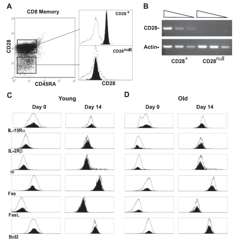
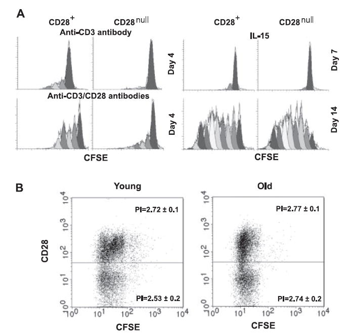
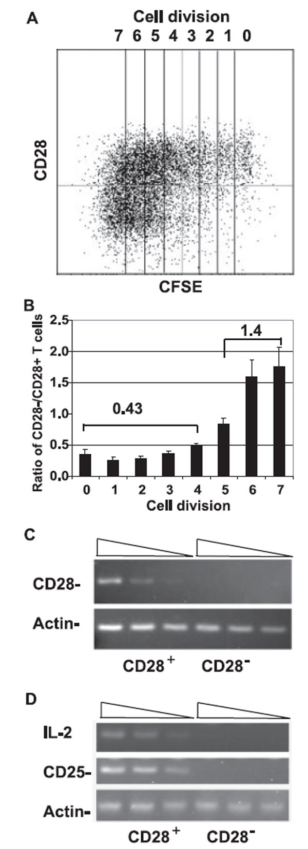
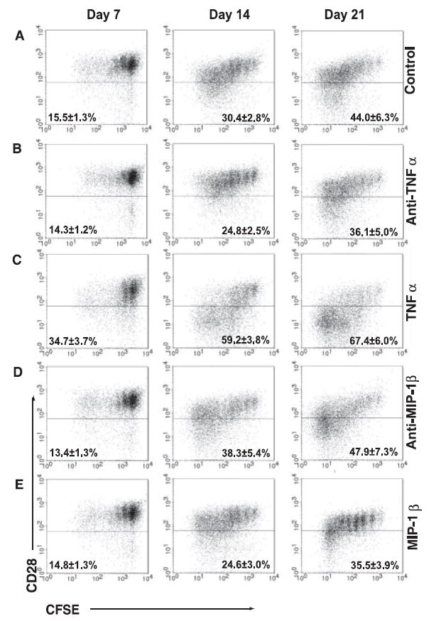
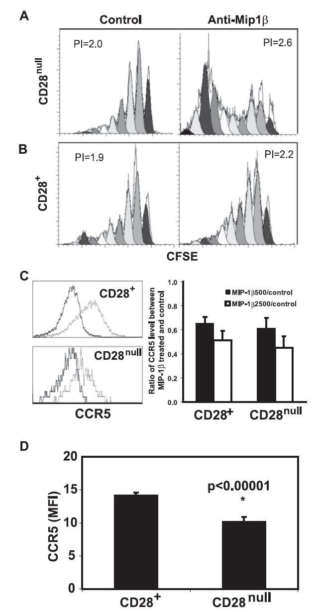
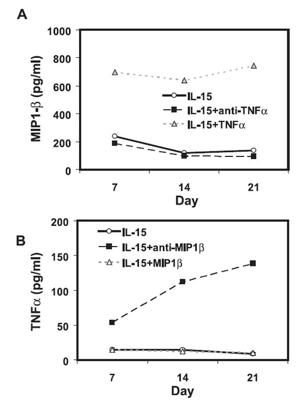
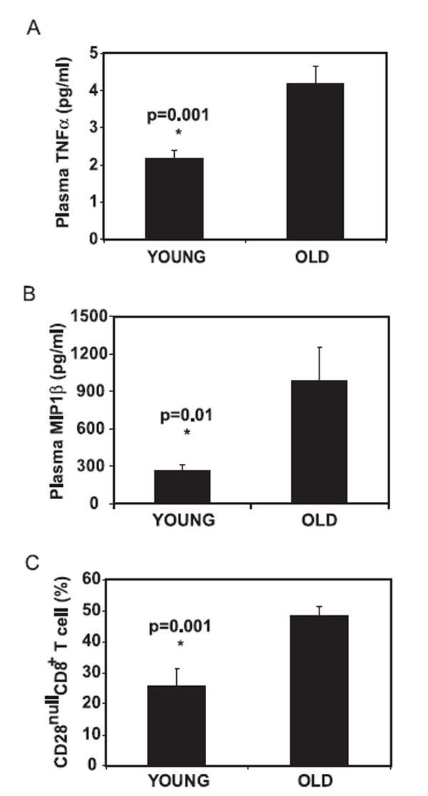
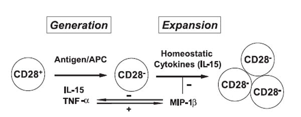
Accumulation of CD28(null)CD8+ T cells and the defects of these cells in response to antigenic stimulation are the hallmarks of age-associated decline of T cell function. However, the mechanism of these age-associated changes is not fully understood. In this study, we report an analysis of the growth of human CD28(null) and CD28+CD8+ memory T cells in response to homeostatic cytokine IL-15 in vitro. We showed that 1) there was no proliferative defect of CD28(null)CD8+ memory T cells in response to IL-15 compared with their CD28+ counterparts; 2) stable loss of CD28 expression occurred in those actively dividing CD28+CD8+ memory T cells responding to IL-15; 3) the loss of CD28 was in part mediated by TNF-alpha that was induced by IL-15; and 4) CCL4 (MIP-1beta), also induced by IL-15, had a significant inhibitory effect on the growth of CD28(null) cells, which in turn down-regulated their expression of CCL4 receptor CCR5. Together, these findings demonstrate that CD28(null)CD8+ memory T cells proliferate normally in response to IL-15 and that IL-15 and its induced cytokines regulate the generation and growth of CD28(null)CD8+ T cells, suggesting a possible role of IL-15 in the increase in CD28(null)CD8+ T cells that occurs with aging.


Conflict of interest statement

Disclosures The authors have no financial conflict of interest.

Figures

FIGURE 1
FIGURE 1
IL-15 induced similar cellular responses between CD28null and CD28+CD8+ memory phenotype T cells in vitro. A, Isolation of CD28null and CD28+CD8+ memory phenotype T cells (filled). Negative control (line) is stained with IgG control Ab. B, CD28 and β-actin gene expressions of freshly isolated CD28null and CD28+CD8+ memory phenotype T cells. A total of 39 cycles of PCR were performed to amplify CD28 and 33 cycles of PCR were performed to amplify β-actin. A serial 4-fold dilution of cDNA was used. C, Expression of IL-15 receptors and apoptosis and survival markers of CD28null (shaded) and CD28+ (line) CD8+ T cells of young donors before and after treatment with IL-15 for 14 days are shown in the histogram. FACS analyses were done on days 0 and 14 after IL-15 treatment by FACS with specific Abs. D, Expression of IL-15 receptors and apoptosis and survival markers of CD28null (filled) and CD28+ (line) CD8+ T cells of old donors before and after treatment with IL-15 for 14 days in are shown in the histogram. In both C and D, representative data are presented from four independent donors. The mean age for young donors is 23 ± 2 and for old donors it is 82 ± 2.
FIGURE 2
FIGURE 2
A, Cell division profiles of CD28null and CD28+CD8+ memory T cells stimulated with anti-CD3 and anti-CD3/CD28 Abs at day 4. CFSE was labeled at day 0 and collected at day 4 by FACScan. Cell division profiles of CD28null and CD28+CD8+ memory T cells stimulated with IL-15 at days 7 and 15. Data were collected by FACScan. A representative histogram is shown from six independent donors prepared by Modfit software. B, Cell division profiles of IL-15-treated CD28null and CD28+CD8+ T cells isolated from young and old donors at day 14. CFSE was labeled at day 0 and data were collected at day 14 by FACScan. Representative figures are shown from eight independent donors (four young donors and four old donors) prepared by CellQuest Pro.
FIGURE 3
FIGURE 3
Loss of CD28 expression in CD28+CD8+ memory phenotype T cells after IL-15 treatment. A, Loss of CD28 expression in frequently dividing CD28+CD8+ memory T cells. Representative data of 11 independent experiments are shown. Freshly sorted CD28+CD8+ memory T cells were labeled with CFSE and cultured in the presence of 50 ng/ml IL-15 for 15 days. Cells were collected on days 5, 10, and 15 and CD28 expressions were analyzed by FACScan. B, The quantitative analysis of loss of CD28 expression in CD28+CD8+ memory phenotype T cells as a function of cell division. The ratios of CD28null to CD28+CD8+ memory T cells of each cell division are presented as mean ± SEM (n = 11). The mean ratios of CD28null/CD28+CD8+ T cells that underwent fewer than four and more than five cell divisions were 0.43 and 1.4, respectively. C, Lack of CD28 transcription in IL-15-generated CD28nullCD8+ memory T cells. CD8 memory T cells were cultured under IL-15 for 14 days before being sorted into CD28null and CD28+ subsets. CD28 expression and β-actin expression were analyzed by 39 cycles and 33 cycles of real-time RT-PCR, respectively. A 4-fold serial dilution of cDNA was applied in the analysis. D, Lack of IL-2 and IL-2Rα (CD25) expression in IL-15-cultured CD28nullCD8+ memory T cells after stimulation. Cells were stimulated with anti-CD3/CD28 beads for 16 h. Real-time RT-PCR was performed with 4-fold serial-diluted cDNA. Forty cycles of PCR were performed to amplify IL-2 and 35 cycles of PCR were performed to amplify CD25 and β-actin.
FIGURE 4
FIGURE 4
IL-15-induced TNF-α and MIP-1β influence the number of CD28null cells in CD28+CD8+ memory T cells. A, Generation of CD28null cells from CD28+CD8+ memory T cells after IL-15 treatment. CD28+CD8+ memory T cells isolated from cell sort were labeled with CFSE and cultured in the presence of IL-15. B, Reduction of CD28null cells by blocking TNF-α with Ab in IL-15-treated CD28+CD8+ memory T cells. Anti-TNF-α treatment (20 μg/ml) was added along with IL-15 to freshly sorted CD28+CD8+ memory T cells. C, Increase of CD28null cells by supplement of TNF-α in IL-15-treated CD28+CD8+ memory T cells. rTNF-α (200 ng/ml) was added along with IL-15 to freshly sorted CD28+CD8+ memory T cells. D, Increase of CD28null cells in the presence of anti-MIP-1β-neutralizing Ab in IL-15-treated CD28+CD8+ memory T cells. Anti-MIP-1β (6 μg/ml) was added along with IL-15 to sorted CD28+CD8+ memory T cells. E, Reduction of CD28null cells in the presence of rMIP-1β. rMIP-1β (500 ng/ml) was added along with IL-15. In all culture conditions, cells were collected at days 7, 14, and 21 and the rate of cell division and CD28 expression were analyzed by FACScan. The representative plots and the mean ± SEM from 5 to 13 independent donors are shown.
FIGURE 5
FIGURE 5
Inhibitory effect of MIP-1β on proliferation of CD28nullCD8+ memory T cells. CD28+CD8+ memory T cells were cultured for 14 days in the presence of IL-15 and sorted into CD28null and CD28+ subsets. Both subsets were labeled with CFSE and cultured with 50% fresh medium and 50% pooled conditional medium collected from IL-15-cultured CD8+ memory T cells of 10 donors. Cells were cultured for 14 days and analyzed by FACScan and software ModFit. A, Anti-MIP-1β treatment had a significant inhibitory effect on proliferation of CD28nullCD8+ memory T cells. A representative histogram is shown. B, Anti-MIP-1β treatment had a less inhibitory effect on proliferation of CD28+CD8+ memory T cells. A representative histogram is shown. The data in A and B were derived from 10 independent donors. C, Down-regulation of CCR5 expression in CD28null and CD28+CD8+ memory T cells in vitro after exposure to rMIP-1β at 500 ng/ml and 2500 ng/ml for 7 days. On the left, histograms of CCR5 expression levels between rMIP-1β treated (2500 ng/ml, bold line) and control (light line) for both CD28null and CD28+CD8+ T cells. On the right, the ratios of CCR5 expression between rMIP-1β treated and untreated CD8 subsets were presented as mean ± SE (n = 4). D, Decrease of CCR5 expression in CD28nullCD8+ T cells ex vivo. The levels of CCR5 on both CD28null and CD28+CD8+ T cells were analyzed by FACSCalibur, and data were presented as mean ± SEM (n = 65).
FIGURE 6
FIGURE 6
Production of TNF-α and MIP-1β by IL-15-treated CD8+ memory T cells in vitro. A, The level of TNF-α is correlated with the level of MIP-1β in the supernatant of CD28+CD8+ memory T cells. The concentrations of MIP-1β in the supernatant of sorted CD28+CD8+ memory T cells that were cultured with IL-15 in the presence of either anti-TNF-α Ab or rTNF-α were determined by BioPlex assay. The mean of five donors is shown. B, MIP-1β negatively regulates level of TNF-α in CD28+CD8+ memory T cells. The concentrations of TNF-α in the supernatant of sorted CD28+CD8+ memory T cells that were cultured with IL-15 in the presence of either anti-MIP-1β Ab or rMIP-1β were determined by BioPlex assay. The mean of seven donors is shown.
FIGURE 7
FIGURE 7
Levels of TNF-α and MIP-1β in peripheral blood. A, Increased concentration of TNF-α. B, Increase of MIP-1β in blood plasma in old donors. C, Increase of the percentage of CD28nullCD8+ memory T cells with age. Whole blood was collected from young (age <34, n = 15) and old (age >70, n = 59) donors. Plasma was isolated and cytokine level was measured by BioPlex and LincoPlex protein array systems, and the percentage of CD28nullCD8+ memory T cells was determined by FACS analysis.
FIGURE 8
FIGURE 8
A two-step model of the accumulation of CD28nullCD8+ memory T cells with age. 1) Stable down-regulation of CD28 expression occurs in CD28+CD8+ memory T cells induced by either repeated antigenic stimulation or cytokines such as IL-15 and TNF-α and 2) proliferation of CD28null CD8+ memory T cells is induced by homeostatic cytokine (IL-15) but is also impeded by other negative growth regulators such as MIP-1β. The cytokines acting to enhance or inhibit generation and expansion of CD28nullCD8+ memory T cells are coregulated, such as the feedback regulation of TNF-α and MIP-1β, to reach a homeostatic balance. With age, the net effects for increase of CD28nullCD8+ memory T cells prevail.

References

    1. Acuto O, Michel F. CD28-mediated co-stimulation: a quantitative support for TCR signalling. Nat Rev Immunol. 2003;3:939–951. - PubMed
    1. Riley JL, June CH. The CD28 family: a T cell rheostat for therapeutic control of t cell activation. Blood. 2005;105:13–21. - PubMed
    1. Azuma M, Phillips JH, Lanier LL. CD28− T lymphocytes: antigenic and functional properties. J Immunol. 1993;150:1147–1159. - PubMed
    1. Posnett DN, Sinha R, Kabak S, Russo C. Clonal populations of T cells in normal elderly humans: the T cell equivalent to “benign monoclonal gammapathy”. J Exp Med. 1994;179:609–618. - PMC - PubMed
    1. Vallejo AN, Weyand CM, Goronzy JJ. T-cell senescence: a culprit of immune abnormalities in chronic inflammation and persistent infection. Trends Mol Med. 2004;10:119–124. - PubMed