. 2006 Nov 15;20(22):3084-8.
doi: 10.1101/gad.402806.

MtHAP2-1 is a key transcriptional regulator of symbiotic nodule development regulated by microRNA169 in Medicago truncatula

Affiliations

MtHAP2-1 is a key transcriptional regulator of symbiotic nodule development regulated by microRNA169 in Medicago truncatula

Jean-Philippe Combier et al. Genes Dev. .

Abstract

In the model legume Medicago truncatula, we identified a new transcription factor of the CCAAT-binding family, MtHAP2-1, for which RNA interference (RNAi) and in situ hybridization experiments indicate a key role during nodule development, possibly by controlling nodule meristem function. We could also show that MtHAP2-1 is regulated by microRNA169, whose overexpression leads to the same nodule developmental block as MtHAP2-1 RNAi constructs. The complementary expression pattern of miR169 and MtHAP2-1 and the phenotype of miR169-resistant MtHAP2-1 nodules strongly suggest, in addition, that the miR169-mediated restriction of MtHAP2-1 expression to the nodule meristematic zone is essential for the differentiation of nodule cells.


Figures
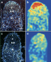
Figure 1.
Figure 1.
MtHAP2-1 expression is nodule specific and restricted to the meristematic-zone mRNA in situ hybridization of MtHAP2-1 in nitrogen-fixing nodules. Sections are hybridized with an antisense MtHAP2-1 probe (a,b) or a control sense MtHAP2-1 probe (c,d). a and c are dark-field microscopy images, to visualize silver grains corresponding to MtHAP2-1 mRNA localization. Nodule zones are indicated according to Vasse et al. (1990) (I, meristematic zone; II, infection zone; II–III, interzone; III, nitrogen fixation zone). b and d are color density maps performed using Image-Pro plus and Diatrack softwares. Bar, 100 μm.
Figure 2.
Figure 2.
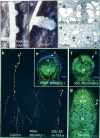
Altered nodulation phenotypes of MtHAP2-1 RNAi and 35S:MtmiR169-a roots. (a,b) Nodule development on roots transformed with either an MtHAP2-1 RNAi construct (a) or a control construct (empty vector) (b). Pictures were taken 46 d post-Rhizobium inoculation (d.p.i.). (c,d) Electron microscopy images of the meristematic zone of MtHAP2-1 RNAi (c) and control (d) nodules. (V) Vacuoles. Cells of the meristematic region of MtHAP2-1 RNAi nodules already start to loose meristematic characteristics at 10 d.p.i. (c), while they are still fully functional in control nodules at 27 d.p.i. (d). Bar, 5 μm. (e–g) Light microscopic images of longitudinal sections of 20-d-old nodules, using an overlay of two fluorescence images. Note the absence of normal nodule zonation and the abnormal closed endodermis (E) in MtHAP2-1 RNAi (e) and Mtmir169-a-overexpressing (f) nodules, compared with control (empty vector) (g) nodules, indicating nodule developmental arrest. Bar, 100 μm. (h) Growth of Rhizobium-inoculated plants after 40 d in the absence of mineral nitrogen reveals a deficient nitrogen fixation phenotype for MtHAP2-1 RNAi and 35S:Mtmir169-a plants compared with control plants.
Figure 3.
Figure 3.
A miR169-a precursor induces cleavage of MtHAP2-1 mRNA. (a) Schematic representation of the MtHAP2-1 gene with intron (line)–exon (boxes) structure and position of the two putative miR169 recognition sites (dotted lines) in the 3′ UTR of the gene. Alignment of miR169 with the two miR169 recognition sites in MtHAP2-1; conserved nucleotides are indicated in bold on the MtHAP2-1 sequence. Arrow shows predominant cleavage site revealed by 5′ RACE–PCR. Underlined nucleotides are those changed by site-directed mutagenesis in the miR169-resistant MtHAP2-1 mutant (miR169-R MtHAP2-1). (b) Most probable stem-loop structure of MtmiR169-a, indicating position of mature miR169 (line) and complementary miR169* (dotted line) sequences.
Figure 4.
Figure 4.
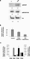
miR169 cleaves MtHAP2-1 in planta and MtHAP2-1 and the MtmiR169-a are oppositely regulated during nodulation. (a) Northern blot analysis of mature 21-bp miR169 expression in transgenic roots transformed with either empty vector control (C) or the 35S:MtmiR169-a constructs (miR). (b) Real-time RT–PCR analysis of MtHAP2-1 expression in MtHAP2-1 RNAi and 35S:MtmiR169-a compared with empty vector controls. Results are expressed as a percentage of the expression in control roots. (c) Real-time RT–PCR analysis of the expression pattern of MtmiR169-a (black boxes) and MtHAP2-1 (striped boxes) during nodule development. Relative expression levels are expressed in arbitrary units; the scale for MtmiR169-a is on the left while the scale for MtHAP2-1 is on the right.
Figure 5.
Figure 5.
MtmiR169-a expression pattern in mature nodules and effects of a MtHAP2-1 miR169-resistant construct on nodule development. (a,b) Expression analysis of MtmiR169-a in 14-d-old nodules revealed by promoter:GUS analysis. b is a magnification of the distal part of the nodule depicted in a, showing the expression of MtmiR169-a in the infection zone (II) and the absence of promoter activity in the meristematic region (I) of the nodule. Description of nodule zones is according to Vasse et al. (1990). Bar, 100 μm. (c–d) Images of longitudinal sections through 20-d-old nodules are an overlay of fluorescence images acquired with two different wavelengths, showing nodule development on transgenic roots expressing either a miR169-resistant MtHAP2-1 (miR169 R-HAP2-1) gene under the control of its own promoter (c), or an empty vector control (control) (d). Bar, 100 μm.

References

    1. Ardourel, M., Demont, N., Debelle, F., Maillet, F., de Billy, F., Prome, J.C., Denarie, J., Truchet, G. Rhizobium meliloti lipooligosaccharide nodulation factors: Different structural requirements for bacterial entry into target root hair cells and induction of plant symbiotic developmental responses. Plant Cell. 1994;6:1357–1374. - PMC - PubMed
    1. Ben-Naim, O., Eshed, R., Parnis, A., Teper-Bamnolker, P., Shalit, A., Coupland, G., Samach, A., Lifschitz, E. The CCAAT binding factor can mediate interactions between CONSTANS-like proteins and DNA. Plant J. 2006;46:462–476. - PubMed
    1. Bhattacharya, A., Deng, J.M., Zhang, Z., Behringer, R., de Crombrugghe, B., Maity, S.N. The B subunit of the CCAAT box binding transcription factor complex (CBF/NF-Y) is essential for early mouse development and cell proliferation. Cancer Res. 2003;63:8167–8172. - PubMed
    1. Boisson-Dernier, A., Andriankaja, A., Chabaud, M., Niebel, A., Journet, E.P., Barker, D.G., de Carvalho-Niebel, F. MtENOD11 gene activation during rhizobial infection and mycorrhizal arbuscule development requires a common AT-rich-containing regulatory sequence. Mol. Plant Microbe Interact. 2005;18:1269–1276. - PubMed
    1. El Yahyaoui, F., Kuster, H., Ben Amor, B., Hohnjec, N., Pühler, A., Becker, A., Gouzy, J., Vernié, T., Gough, C., Niebel, A., et al. Expression profiling in Medicago truncatula identifies more than 750 genes differentially expressed during nodulation, including many potential regulators of the symbiotic program. Plant Physiol. 2004;136:3559–3576. - PMC - PubMed

Publication types

MeSH terms

LinkOut - more resources