. 2006 Nov 15;20(22):3104-16.
doi: 10.1101/gad.1478906.

The S. cerevisiae Rrm3p DNA helicase moves with the replication fork and affects replication of all yeast chromosomes

Affiliations

The S. cerevisiae Rrm3p DNA helicase moves with the replication fork and affects replication of all yeast chromosomes

Anna Azvolinsky et al. Genes Dev. .

Abstract

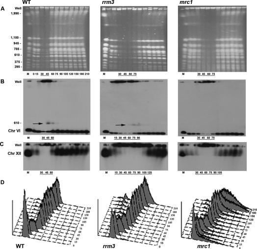
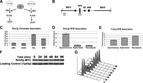
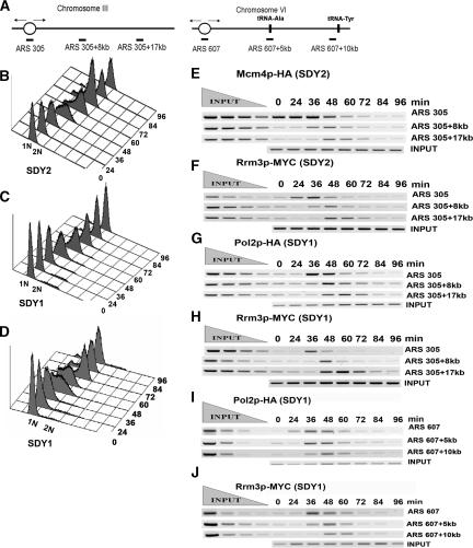
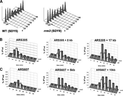
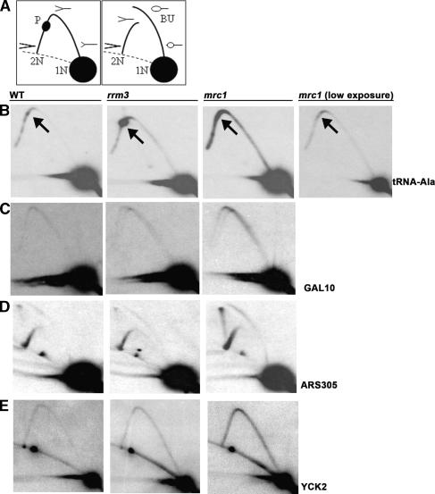
The Saccharomyces cerevisiae DNA helicase Rrm3p is needed for normal fork progression through >1000 discrete sites scattered throughout the genome. Here we show that replication of all yeast chromosomes was markedly delayed in rrm3 cells. Delayed replication was seen even in a region that lacks any predicted Rrm3p-dependent sites. Based on the pattern of replication intermediates in two-dimensional gels, the rate of fork movement in rrm3 cells appeared similar to wild-type except at known Rrm3p-dependent sites. These data suggest that although Rrm3p has a global role in DNA replication, its activity is needed only or primarily at specific, difficult-to-replicate sites. By the criterion of chromatin immunoprecipitation, Rrm3p was associated with both Rrm3p-dependent and -independent sites, and moved with the replication fork through both. In addition, Rrm3p interacted with Pol2p, the catalytic subunit of DNA polymerase epsilon, in vivo. Thus, rather than being recruited to its sites of action when replication forks stall at these sites, Rrm3p is likely a component of the replication fork apparatus.


Figures

Figure 1.
Figure 1.
Replication of all chromosomes is delayed in rrm3 cells. All time points are indicated below the ethidium bromide-stained wild-type (WT) gel. For ease of viewing, only time points where chromosomes are replicating are indicated below all subsequent gels and Southern blots. (A) Ethidium bromide-stained PFGs of chromosomal-sized DNA molecules from wild-type, rrm3, and mrc1 strains. Chromosomal markers (M) are indicated on the left in kilobases. (B) PFGs were visualized by Southern hybridization with a chromosome VI probe (HIS2). Arrows indicate a form of chromosome VI detected in late S phase that behaves like a linear dimer. The membrane in B was stripped and reprobed sequentially for chromosome XIV (ARS1414 probe; Supplementary Fig. 1) and chromosome XII (rDNA probe; C). (D) Flow-cytometric profiles of synchronized cultures.
Figure 2.
Figure 2.
Delayed replication of segments of chromosome XII and XIV in rrm3 cells. The DNA samples prepared for Figure 1 were digested with NotI and then analyzed by PFG and Southern hybridization. Symbols are the same as in Figure 1. The positions of NotI sites in chromosomes XII (A) and XIV (C) are indicated. Black circles represent centromeres. Time points are labeled as in Figure 1. After separation by PFGs, NotI-digested DNA was transferred to membranes and hybridized sequentially with probes that detect the 107-kb telomeric NotI fragment from chromosome XII (AYTI probe; A), the penultimate 101-kb NotI fragment from the right arm of chromosome XII (CRN1 probe; Supplementary Fig. 2B), the entire rDNA array (rDNA probe; B), and a 129-kb NotI fragment on chromosome XIV (ARS1414 probe; C).
Figure 3.
Figure 3.
Rrm3p associates with Rrm3p-dependent and -independent sites in a cell cycle-dependent manner. (A) Schematic of cell-mixing experiment used for C and D. The A+ strain contains a portion of ADH4 that is absent from the no-tag control and Rrm3p-MYC strains. (B) Schematic of a single 9.1-kb rDNA repeat. Bars indicate positions of PCR-amplified sequences used for D (RFB) and E (RFB, 35S). (C) DNA immunoprecipitated from the Rrm3p-MYC strain was PCR-amplified for 23 cycles using primers for the RFB and for 28 cycles using primers for the single-copy TEL, ARO, or ADH sequences. Intensities of amplified fragments were quantified by densitometric analysis. The A+ sequence, which is absent from experimental strains, was used for background normalization. Fold enrichment ± standard deviations are indicated here and in D and E. (D) DNA was immunoprecipitated from asynchronous, G1 phase, and G2/M phase Rrm3p-MYC cells and amplified using 23 cycles of multiplex PCR with primers specific to the RFB. The amount of RFB sequence in the immunoprecipitate was normalized to the background intensity of the A+ sequence. (E) DNA was immunoprecipitated as described for D from a Fob1p-MYC strain except that the amount of RFB sequence in the immunoprecipitate was normalized to a sequence from within the 35S-encoding rDNA. (F) Rrm3p-MYC abundance is constant throughout the cell cycle. Synchronized Rrm3p-MYC cells were collected at the indicated time points and analyzed for Rrm3p-MYC abundance after separation by SDS-PAGE and Western blotting with MYC antibody and subsequently with Tpd3 antibody. (G) Flow-cytometric profile of synchronized cells used in F.
Figure 4.
Figure 4.
Rrm3p loads onto origins at the beginning of S phase and migrates with the replication fork through both Rrm3p-dependent and -independent sites. (A) Diagram of the ARS305 region on chromosome III (left) and the ARS607 region on chromosome VI (right). The oval indicates the origin of DNA replication. Vertical bars indicate the positions of two tRNA genes to the right of ARS607. Horizontal bars indicate the positions of the segments amplified by PCR. (B-D) Flow-cytometric profiles for strains used for E and F (SDY2 cells, express Mcm4p-HA and Rrm3p-MYC), G and H (SDY1 cells, express Pol2p-HA and Rrm3p-MYC), and I and J and Supplementary Figure 3 (SDY1 cells, express Pol2p-HA and Rrm3p-MYC). (E,F) SDY2 cells were arrested in late G1 phase and released synchronously into the cell cycle at 18°C. Samples were collected for flow cytometry, ChIP, and Western blot analysis at 12-min intervals. Formaldehyde-fixed cells were immunoprecipitated with anti-HA (Mcm4p, E) or anti-MYC (Rrm3p, F) antibodies. DNA was purified, PCR-amplified, and resolved on a 2.8% ethidium bromide agarose gel. Each primer set was used to amplify both input and immunoprecipitated DNA but only one representative input reaction per site is shown. (G,H). Synchronized SDY1 cells were processed for ChIP and analyzed for association of Pol2p-HA (G) and Rrm3p-MYC (H) with the ARS305 region as described for E and F. (I,J) Synchronized SDY1 cells were processed for ChIP and analyzed for association of Pol2p-HA (I) and Rrm3p-MYC (J) with the ARS607 region as described for E and F.
Figure 5.
Figure 5.
Rrm3p associates with Pol2p during S phase. Protein extracts were prepared from mid-S-phase cultures of strains SDY1 (A) and SDY2 (B), treated with DNase I (+) or not (−), and immunoprecipitated (IP) with either anti-C-MYC Agarose-conjugated beads or anti-HA Agarose-conjugated beads. Input and IP samples were separated on SDS-PAGE gels, and analyzed by Western blotting. Blots were probed with anti-HA (top) or anti-MYC (bottom).
Figure 6.
Figure 6.
Pol2p migrates more slowly in the absence of Rrm3p through Rrm3p-dependent sites. Strains SDY5 (expresses Pol2p-MYC) and SDY8 (rrm3 expresses Pol2p-MYC) were synchronized and processed for ChIP as described in Figure 4. (A) Flow-cytometric profiles for synchronized SDY5 and SDY8. (B) The association of Pol2p-MYC with the ARS305 region (diagrammed in Fig. 4A) with (white bars) and without (gray bars) Rrm3p was examined. (C) DNA from the same samples used in B was analyzed for the association of Pol2p-HA with the ARS607 region, which contains two Rrm3p-dependent sites (diagrammed in Fig. 4A, right), with (white bars) and without (gray bars) Rrm3p. In B and C, the intensities of amplified fragments were quantified by densitometric analysis. The percentage of input DNA precipitated (% IP’ed) is graphed for one of the two independent experiments performed with each strain.
Figure 7.
Figure 7.
Abundance of replication intermediates in wild-type (WT), rrm3, and mrc1 strains. (A) Schematics of replication intermediates as visualized in 2D gels. (1N) Nonreplicating fragment; (2N) almost fully replicated fragment right before sister chromatids separate; (P) replication pause; (BU) bubble-shaped replication intermediates. Dotted lines indicate arc formed by linear DNA molecules. (B–E) DNA from glucose-grown asynchronous cells from the indicated strains was restriction enzyme-digested, separated on 2D gels, and analyzed by Southern blotting with the indicated probes (restriction enzymes and hybridization probes are noted in parentheses). (B) tRNAA (BglII, HIS2); arrows indicate the position of the tRNA gene on the arc of replication intermediates; a lower exposure of the sample from the mrc1 strain is shown to provide better visualization of fork movement through the tRNA gene. (C) GAL10 (PvuII, GAL10). (D) ARS305 (EcoRV, ARS305). (E) YCK2 (BglII, YCK2).

Similar articles

Cited by

References

    1. Admire, A., Shanks, L., Danzl, N., Wang, M., Weier, U., Stevens, W., Hunt, E., Weinert, T. Cycles of chromosome instability are associated with a fragile site and are increased by defects in DNA replication and checkpoint controls in yeast. Genes & Dev. 2006;20:159–173. - PMC - PubMed
    1. Alcasabas, A., Osborn, A., Bachant, J., Hu, F., Werler, P., Bousset, K., Furuya, K., Diffley, J., Carr, A., Elledge, S. Mrc1 transduces signals of DNA replication stress to activate Rad53. Nat. Cell Biol. 2001;3:958–965. - PubMed
    1. Aparicio, O.M., Weinstein, D.M., Bell, S.P. Components and dynamics of DNA replication complexes in S. cerevisiae: Redistribution of MCM proteins and Cdc45p during S phase. Cell. 1997;91:59–69. - PubMed
    1. Barry, J., Alberts, B. A role for two DNA helicases in the replication of T4 bacteriophage DNA. J. Biol. Chem. 1994;269:33063–33068. - PubMed
    1. Bedinger, P., Hochstrasser, M., Jongeneel, C., Alberts, B. Properties of the T4 bacteriophage DNA replication apparatus: The T4 dda DNA helicase is required to pass a bound RNA polymerase molecule. Cell. 1983;34:115–123. - PubMed

Publication types

MeSH terms