. 2007 Jan;6(1):48-59.
doi: 10.1128/EC.00318-06. Epub 2006 Nov 17.

Vesicular polysaccharide export in Cryptococcus neoformans is a eukaryotic solution to the problem of fungal trans-cell wall transport

Affiliations

Vesicular polysaccharide export in Cryptococcus neoformans is a eukaryotic solution to the problem of fungal trans-cell wall transport

Marcio L Rodrigues et al. Eukaryot Cell. 2007 Jan.

Abstract

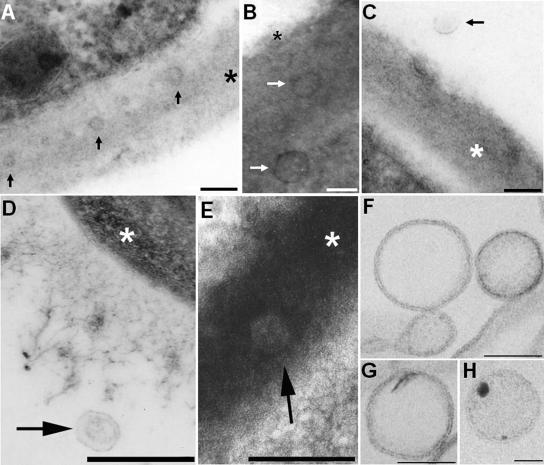
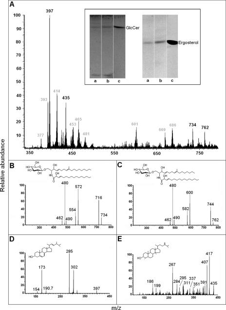
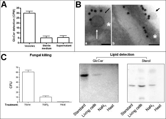
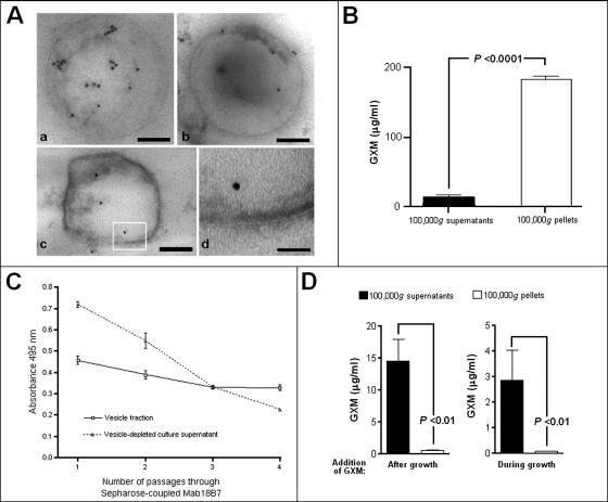
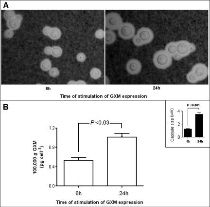
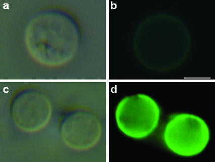
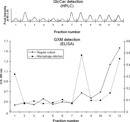
The mechanisms by which macromolecules are transported through the cell wall of fungi are not known. A central question in the biology of Cryptococcus neoformans, the causative agent of cryptococcosis, is the mechanism by which capsular polysaccharide synthesized inside the cell is exported to the extracellular environment for capsule assembly and release. We demonstrate that C. neoformans produces extracellular vesicles during in vitro growth and animal infection. Vesicular compartments, which are transferred to the extracellular space by cell wall passage, contain glucuronoxylomannan (GXM), a component of the cryptococcal capsule, and key lipids, such as glucosylceramide and sterols. A correlation between GXM-containing vesicles and capsule expression was observed. The results imply a novel mechanism for the release of the major virulence factor of C. neoformans whereby polysaccharide packaged in lipid vesicles crosses the cell wall and the capsule network to reach the extracellular environment.


Figures

FIG. 1.
FIG. 1.
TEM of vesicles in acapsular (A to C) and encapsulated (D to H) C. neoformans cells. The occurrence of vesicles in association with the cell wall of acapsular cryptococci (A and B) or in the extracellular environment (C) is evident after in vitro growth. Vesicle-like structures were also observed in the lung following murine pulmonary infection (D and E). Putative vesicles near the edge of the capsule (D) or in the cryptococcal cell wall (E) were observed 2 h after infection. Bars, 100 nm (A to C) and 500 nm (D and E). Arrows point to vesicles, and asterisks are on the cryptococcal cell wall. (F to H) The pellets obtained by ultracentrifugation were isolated by differential centrifugation, purified from GXM by affinity chromatography, and analyzed by TEM. Extracellular vesicles with bilayered membranes and different profiles of electron density were observed. Bars, 100 nm (F and G) and 50 nm (H).
FIG. 2.
FIG. 2.
Lipid analysis of C. neoformans vesicles. (A) Lipid extracts were prepared as described in Materials and Methods and analyzed by ESI-MS and HPTLC. Vesicle lipids with migration rates corresponding to GlcCer and ergosterol standards (c) were detected by HPTLC (inset) in preparations from both encapsulated (a) and acapsular (b) C. neoformans cells. ESI-MS analysis demonstrated the presence of molecular ions corresponding to GlcCer (734 and 762) and sterols (435 and 397) besides other still-unidentified molecules (m/z values in gray). Fragmentation analysis of molecular ions at m/z 734 (B) and 762 (C) revealed ionized species with m/z values typical of lithiated fungal cerebrosides. Ceramide structures with C16 (structure in panel B) and C18 (structure in panel C) fatty acids were detected. Fragmentation analysis of molecular ions at m/z 397 (D) and 435 (E) revealed the presence of ergosterol (structure in panel D) and 4,14-dimethylergosta-24(241)-en-3β-ol (structure in panel E), an obtusifoliol-like structure.
FIG. 3.
FIG. 3.
Vesicle production requires cell viability. (A) The addition of radioactive ceramide precursors to the culture medium results in the detection of significant levels of radioactive GlcCer in vesicle preparations. In 100,000 × g pellets from sterile medium and in 100,000 × g supernatants of grown cells, significant levels of radioactive GlcCer were not detected (P < 0.001). (B) GlcCer-containing vesicles are apparently transferred from the plasma membrane to the cell wall (a) and then secreted (b). Scale bars represent 100 nm. Arrows point to vesicles, and asterisks are on the cryptococcal cell wall. (C) Killing of cryptococci with sodium azide or heating demonstrates a decrease in cell viability of 83% (sodium azide) and 99% (heat), as determined by comparison with CFU counts obtained with untreated yeasts. Lipid analysis of 100,000 × g fractions of the supernatants obtained after fungal killing revealed the detection of GlcCer in fractions from living but not heat- or azide-treated cells. Compounds with migration rates corresponding to an ergosterol standard were detected in 100,000 × g fractions from supernatants of living and azide-treated cells but not in preparations from heat-killed cryptococci. CPM, counts per minute.
FIG. 4.
FIG. 4.
GXM is present in purified vesicles. (A) Immunogold labeling of purified vesicles with MAb 18B7 revealed a preferential intravesicular distribution of GXM, as observed in different vesicular bodies (a to c). A higher magnification of the boxed area shown in panel c demonstrates the occurrence of bilayered membrane (d). Bars, 150 (a), 180 (b), 120 (c), and 30 (d) nm. (B) The presence of GXM inside the vesicles was confirmed by polysaccharide detection in purified 100,000 × g fractions from culture supernatants (white bar). Supernatants obtained from these suspensions were assayed as a control of vesicle integrity, and in fact, GXM was detected at low levels in these preparations (black bar), suggesting that some of these structures may be disrupted. (C) GXM content of vesicle suspensions and vesicle-depleted supernatants after serial passage through a column composed of Sepharose-bound MAb 18B7. Sequential passages of vesicle-depleted supernatant through this column result in a rapid loss of reactivity, while the GXM content of the vesicular preparation is relatively constant. (D) GXM-containing vesicles are not formed extracellularly, since the addition of the polysaccharide to culture supernatants during or after growth of Cap67 cells is not followed by its detection in purified 100,000 × g fractions.
FIG. 5.
FIG. 5.
Association of capsule expression with supernatant accumulation of GXM-containing vesicles. (A) Capsule expression was higher in yeast cells incubated for 24 h in capsule-inducing media. (B) The profile of GXM detection in ultracentrifugation pellets was very similar to that observed for determinations of capsule size (inset), suggesting a correlation between capsule expression and the production of vesicles containing GXM. GXM concentrations in 100,000 × g fractions under the different conditions of capsule induction were normalized to the number of cells in the culture under each experimental condition.
FIG. 6.
FIG. 6.
Acapsular cells of C. neoformans bind GXM from extracellular vesicles. Cap67 cells were incubated in the presence of purified vesicles and then analyzed by immunofluorescence with MAb 18B7. Control cells, which were not incubated with MAb 18B7, are shown in the upper panels (a and b). Yeast cells that were incubated in the presence of the vesicular preparation reacted strongly with the antibody to GXM, as shown in the lower panels (c and d). The left panels (a and c) represent cryptococci analyzed under differential interference contrast, while the right panels (b and d) show images in the fluorescence mode. Bar, 2 μm.
FIG. 7.
FIG. 7.
Analysis of vesicle fractions obtained after ultracentrifugation and sucrose gradient separation. Fractions from supernatants of regular cultures were extracted with chloroform-methanol mixtures and analyzed by HPLC. In each fraction, a single peak with a retention time (Rt) corresponding to a GlcCer standard was detected. Analysis of the same fractions (solid line) or preparations obtained from infected macrophages (dashed line) by capture ELISA revealed different profiles of GXM distribution, although the polysaccharide was always expressively detected in the region of the gradient presenting the highest density. Lipid and polysaccharide analyses were performed at least three times, always presenting similar profiles. O.D. 405 nm, optical density at 405 nm.

Similar articles

Cited by

References

    1. Barreto-Bergter, E., M. R. Pinto, and M. L. Rodrigues. 2004. Structure and biological functions of fungal cerebrosides. An. Acad. Bras. Cienc. 76:67-84. - PubMed
    1. Casadevall, A., J. Mukherjee, and M. D. Scharff. 1992. Monoclonal antibody based ELISAs for cryptococcal polysaccharide. J. Immunol. Methods 154:27-35. - PubMed
    1. Casadevall, A., W. Cleare, M. Feldmesser, A. Glatman-Freedman, D. L. Goldman, T. R. Kozel, N. Lendvai, J. Mukherjee, L. A. Pirofski, J. Rivera, A. L. Rosas, M. D. Scharff, P. Valadon, K. Westin, and Z. Zhong. 1998. Characterization of a murine monoclonal antibody to Cryptococcus neoformans polysaccharide that is a candidate for human therapeutic studies. Antimicrob. Agents Chemother. 42:1437-1446. - PMC - PubMed
    1. Chang, Y. C., and K. J. Kwon-Chung. 1994. Complementation of a capsule-deficient mutation of Cryptococcus neoformans restores its virulence. Mol. Cell. Biol. 14:4912-4919. - PMC - PubMed
    1. Chen, S. C., L. C. Wright, R. T. Santangelo, M. Muller, V. R. Moran, P. W. Kuchel, and T. C. Sorrell. 1997. Identification of extracellular phospholipase B, lysophospholipase, and acyltransferase produced by Cryptococcus neoformans. Infect. Immun. 65:405-411. - PMC - PubMed

Publication types