. 2007 Jan 12;282(2):986-95.
doi: 10.1074/jbc.M609322200. Epub 2006 Nov 17.

Mechanism of Dun1 activation by Rad53 phosphorylation in Saccharomyces cerevisiae

Affiliations

Mechanism of Dun1 activation by Rad53 phosphorylation in Saccharomyces cerevisiae

Sheng-hong Chen et al. J Biol Chem. .

Abstract

Despite extensive studies, the molecular mechanism of DNA damage checkpoint activation remains incompletely understood. To better dissect this mechanism, we developed an activity-based assay for Dun1, a downstream DNA damage check-point kinase in yeast, using its physiological substrate Sml1. Using this assay, we confirmed the genetic basis of Dun1 activation. Rad53 was found to be directly responsible for Dun1 activation. We reconstituted the activation of Dun1 by Rad53 and found that phosphorylation of Thr-380 in the activation loop of Dun1 by Rad53 is responsible for Dun1 activation. Interestingly, phosphorylation of the evolutionarily conserved Thr-354 in the activation loop of Rad53 is also important for the regulation of Rad53 activity. Thus, this conserved mode of activation loop phosphorylation appears to be a general mechanism for the activation of Chk2 family kinases.


Figures
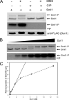
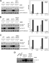
FIGURE 1
FIGURE 1. Analysis of Dun1 activity using Sml1
A, endogenous Dun1 purified from untreated or MMS-treated Dun1-TAF cells was assayed for its kinase activity. Top panel, Coomassie staining of Sml1 after each kinase reaction. Middle panel, autoradiography of Sml1. Bottom panel, anti-FLAG Western blot shows the loading control of Dun1. B, dependence of Sml1 hyperphosphorylation on the concentrations of Dun1 purified from MMS-treated Dun1-TAF cells. A 2-fold increase of Dun1, starting from 0.2 nm, was used to phosphorylate Sml1 (2.5 mm). Upper panel, Coomassie staining of Sml1. Lower panel, autoradiography of Sml1. C, the amount of phosphorylated Sml1 was quantified using scintillation counting. An approximate linear relationship was found between the concentration of Dun1 and the amount of Sml1 phosphorylation when Dun1 concentration is low.
FIGURE 2
FIGURE 2. Analysis of Dun1 activity purified from various mutant backgrounds
A, upper panel, Coomassie staining of Sml1. Middle panel, autoradiography of Sml1. Bottom panel, anti-FLAG Western blot shows the loading control of Dun1 used. A, effect of MEC1 and TEL1 deletion on Dun1 activity. B, effect of RAD9 and MRC1 deletion on Dun1 activity. C, effect of RAD53 and CHK1 deletion on Dun1 activity. D, closer examination of Dun1 activity in various checkpoint mutants, using 5-fold more (10 ng) of Dun1 purified from each mutant.
FIGURE 3
FIGURE 3
A, Coomassie staining of purified recombinant Rad53, including wild type, 4TA mutant, and kinase-dead mutant. B, in vitro activation of Dun1 using Rad53. 1 ng of WT or 4TA mutant Rad53 was used. C, phosphorylation maps of Dun1 revealed that Rad53 is entirely responsible for Dun1 phosphorylation. D, summary of autophosphorylation sites of Dun1 and the Rad53-induced transphosphorylation sites of Dun1 identified using quantitative MS. N/D indicates not determined. # indicates that the corresponding phosphopeptide without Rad53 phosphorylation was not detected; thus the value refers to signal to noise ratio. + indicates the phosphorylated Ser/Thr. * indicates the residue at the +1 position.
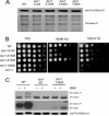
FIGURE 4
FIGURE 4
A, Western blot analysis of the abundance of WT Dun1 and various Dun1 phosphorylation-site mutants with the control of Ponceau S staining of the same. B, UV and hydroxyurea sensitivities of various Dun1 phosphorylation-defective mutants. A serial dilution of various cells was spotted on either HU-containing YPD plate or YPD plate and subjected to UV treatment. C, comparison of the activity of WT, Dun1-KD, and Dun1-T380A for Sml1 hyperphosphorylation.
FIGURE 5
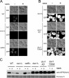
FIGURE 5. GFP-Sml1 abundance in rad9Δ, mrc1Δ, and dun1-T380A mutants
A, Sml1 abundance is analyzed in rad9Δ and mrc1Δ cells. B, GFP-Sml1 abundance in dun1-T380A cells is similar to dun1-KD in response to MMS treatment. C, anti-green fluorescent protein Western blot analysis of GFP-Sml1.
FIGURE 6
FIGURE 6
A, sequence alignments of various Dun1 orthologs, indicating that Thr-380 in the activation loop of Dun1 kinase domain is conserved in the Chk2 family kinases. + indicates the conserved Thr residue; * indicates the residue at the +1 position. B, Sml1 hyperphosphorylation assay using 50 pg of Rad53 and 2 ng of Dun1 purified from rad53Δ cells. Upper panel, Coomassie staining of Sml1. Middle panel, 32P labeling of Sml1. Lower panel, anti-FLAG Western blot detects Dun1. Right panel, scintillation counting of the amount of 32P incorporation of the phosphorylated Sml1. C, UV and hydroxyurea sensitivities of WT, rad53-KD, and rad53-T354A mutant.
FIGURE 7
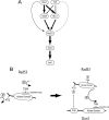
FIGURE 7
A, summary of the roles of various upstream proteins in Dun1 activation. The thicker arrow indicates a larger contribution to Dun1 activation. B, model on Dun1 activation via Rad53 phosphorylation. Rad53 is autophosphorylated on Thr-354, which activates Rad53. Phosphorylated Rad53 also interacts with Dun1 via the FHA domain of Dun1 and the N-terminal phosphorylation cluster of Rad53. This interaction facilitates the phosphorylation of Thr-380 of Dun1 by Rad53, leading to Dun1 activation.

References

    1. Nyberg KA, Michelson RJ, Putnam CW, Weinert TA. Annu. Rev. Genet. 2002;36:617–656. - PubMed
    1. Jackson SP. Curr. Opin. Genet. Dev. 1996;6:19–25. - PubMed
    1. Sanchez Y, Desany BA, Jones WJ, Liu Q, Wang B, Elledge SJ. Science. 1996;271:357–360. - PubMed
    1. Osborn AJ, Elledge SJ. Genes Dev. 2003;17:1755–1767. - PMC - PubMed
    1. Alcasabas AA, Osborn AJ, Bachant J, Hu F, Werler PJ, Bousset K, Furuya K, Diffley JF, Carr AM, Elledge SJ. Nat. Cell Biol. 2001;3:958–965. - PubMed

Publication types

MeSH terms