. 2007 Feb;120(2):230-41.
doi: 10.1111/j.1365-2567.2006.02494.x. Epub 2006 Nov 20.

Increased proteolysis of diphtheria toxin by human monocytes after heat shock: a subsidiary role for heat-shock protein 70 in antigen processing

Affiliations

Increased proteolysis of diphtheria toxin by human monocytes after heat shock: a subsidiary role for heat-shock protein 70 in antigen processing

Barbara S Polla et al. Immunology. 2007 Feb.

Abstract

The expression of heat-shock proteins (hsp) increases after exposure to various stresses including elevated temperatures, oxidative injury, infection and inflammation. As molecular chaperones, hsp have been shown to participate in antigen processing and presentation, in part through increasing the stability and expression of major histocompatibility complex molecules. Heat shock selectively increases human T-cell responses to processed antigen, but does not affect T-cell proliferation induced by non-processed antigens. Here, we have analysed the mechanisms by which stress such as heat shock, and the ensuing hsp over-expression affect the processing of diphtheria toxin (DT) in peripheral blood monocytes. We found that heat shock increased DT proteolysis in endosomes and lysosomes while the activities of the cathepsins B and D, classically involved in DT proteolysis, were decreased. These effects correlated with the heat-shock-mediated increase in hsp 70 expression observed in endosomes and lysosomes. Actinomycin D or blocking anti-hsp 70 antibodies abolished the heat-shock-mediated increase in DT proteolysis. These data indicate that the increased expression of hsp 70 constitutes a subsidiary mechanism that facilitates antigen proteolysis in stressed cells. Confirming these data, presentation by formaldehyde-fixed cells of DT proteolysates that were obtained with endosomes and lysosomes from heat-shocked peripheral blood monocytes showed higher stimulation of T cells than those generated with endosomes and lysosomes from control peripheral blood monocytes.


Figures

Figure 1
Figure 1
HS-induced expression of hsp in human PBM. (a) SDS–PAGE analysis of protein synthesis by PBM after exposure to the indicated temperature. While the expression of inducible hsp 65, hsp 70, hsp 90 and hsp 110 was observed after exposure to 44° and 45°, the decrease in normal protein synthesis only occurred after exposure of PBM to 45°. (b) SDS–PAGE analysis of protein synthesis by PBM after treatment with actinomycin D (AD; 5 μg/ml) added 10 min before HS to parallel cultures and removed at the end of HS. The hsp 65, hsp 70, hsp 90 and hsp 110 induced after exposure to 44° were no longer detectable when actinomycin D had been added, whereas total protein synthesis was unaltered.
Figure 2
Figure 2
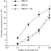
Comparative efficiency to present DT of PBM that have been exposed or not to HS. T lymphocytes (1 × 10⁵) and autologous PBM (3 × 10⁴) exposed or not at 44° for 20 min (in the presence or not of actinomycin D) were cocultured in the presence of increasing concentrations of DT. T-cell proliferation was estimated, as described in the Materials and methods. DT presentation by PBM pre-exposed to HS was more efficient for T-cell stimulation. This effect was abolished in the presence of actinomycin D.
Figure 3
Figure 3
Effect of HS on DT proteolysis by human monocytes. (a) Endosomes (E), lysosomes (L) and cytosol (C) (corresponding to equal cell number) prepared from PBM exposed or not to HS (20 min at 44°) with or without actinomycin D (AD) were incubated for 4 hr at 37° with 125I-labelled DT before being analysed in SDS–PAGE under reducing conditions. DT proteolysis which took place in endosomes and lysosomes (lanes 2 and 3) was increased following HS (lanes 5 and 6). This effect was partly inhibited by actinomycin D (lanes 8 and 9). Lane 1: control DT. (b) When endosomes and lysosomes were exposed to HS after subcellular fractionation (excluding hsp synthesis), elevation of temperature had no effect on DT proteolysis (compare lanes 4 and 5 to lanes 6 and 7). Column markers: H, heavy chain; L, light chain.
Figure 4
Figure 4
Kinetics of DT proteolysis by purified cathepsins B and D. (a) SDS–PAGE analysis in reducing conditions of DT digested by cathepsins B and D, at 15 min, 30 min, and 1, 2, 4 and 6 hr at 37°. Both cathepsins are able to digest DT in a manner similar to subcellular endosomes and lysosomes. (b) Effect of HS on the activity of cathepsins B and D. The activities were estimated in endosomes (E) and lysosomes (L) from cells exposed or not to HS using specific substrates as described in the Materials and methods. (IAA, iodoacetamide; pepA, pepstatine A; H, heavy chain; L, light chain). HS down-regulated both cathepsin's activities in endosomes as well as in lysosomes.
Figure 5
Figure 5
Subcellular localization of hsp following HS. (a) Western blot analysis of hsc and hsp 70 expression following HS (lanes 2); controls are shown in lanes 1. The SPA820 antibody directed against the constitutive hsc 70 cross-reacts with the inducible hsp 70 and recognizes both hsp 70 and hsc 70 (anti-hsc/hsp 70, lane 2 upper panel), whereas the SPA810 antibody only recognizes the inducible hsp 70 (lane 2, lower panel). (b) Western blot analysis of subcellular localization of hsp 70 following HS. HS induced hsp 70 expression in endosomes and lysosomes (lanes 2 and 5). This effect was inhibited by actinomycin D (lanes 3 and 6).
Figure 6
Figure 6
Inhibition of HS increases DT proteolysis by antibodies against hsc/hsp 70. SDS–PAGE analysis of DT proteolysis by endosomes and lysosomes from control or heat-shocked cells, performed for 4 hr at 37° in the presence or absence of antibodies against hsc/hsp 70 (SPA820 Ab). Antibodies for hsc/hsp 70 block the increased proteolysis of DT in lysosomes and endosomes from heat-shocked cells (lanes 4 and 8 compared to lanes 3 and 7). Irrelevant antibodies (Irrel. Ab) had no effect on DT proteolysis (lanes 5 and 9). Lane 1: control DT.
Figure 7
Figure 7
DT proteolysis by endosomes/lysosomes from heat-shocked PBM leads to enhanced T-cell proliferation. DT was incubated for 4, 8 or 24 hr in the presence of endosome/lysosome-enriched fractions from PBM that had been exposed or not to HS. The resulting proteolysates were then presented to autologous T cells by formaldehyde-fixed PBM.
Figure 8
Figure 8
Model for antigen processing under stress conditions. See the text.

References

    1. Rothman JE. Polypeptide chain binding proteins: catalysts of protein folding and related processes in cells. Cell. 1989;59:591–601. - PubMed
    1. Gething MJ, Sambrook J. Protein folding in the cell. Nature. 1992;355(6355):33–45. - PubMed
    1. Liu RY, Li X, Li L, Li GC. Expression of human hsp70 in rat fibroblasts enhances cell survival and facilitates recovery from translational and transcriptional inhibition following heat shock. Cancer Res. 1992;52:3667–73. - PubMed
    1. Jacquier-Sarlin MR, Fuller K, Dinh-Xuan AT, Richard MJ, Polla BS. Protective effects of hsp70 in inflammation. Experientia. 1994;50:1031–8. - PubMed
    1. Germain RN, Margulies DH. The biochemistry and cell biology of antigen processing and presentation. Annu Rev Immunol. 1993;11:403–50. - PubMed

Publication types