. 2007 Feb;27(3):899-911.
doi: 10.1128/MCB.00756-06. Epub 2006 Nov 20.

Abr and Bcr, two homologous Rac GTPase-activating proteins, control multiple cellular functions of murine macrophages

Affiliations

Abr and Bcr, two homologous Rac GTPase-activating proteins, control multiple cellular functions of murine macrophages

Young Jin Cho et al. Mol Cell Biol. 2007 Feb.

Abstract

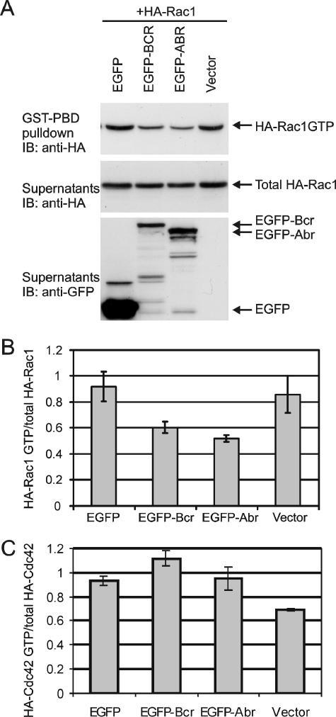
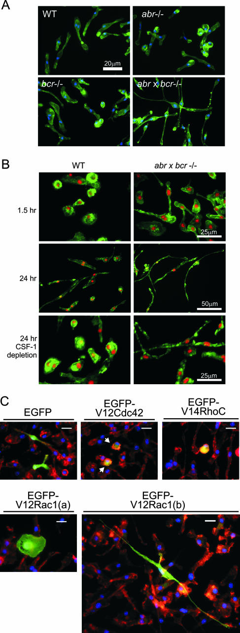
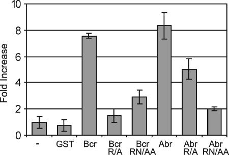
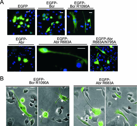
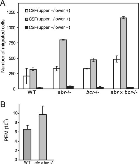
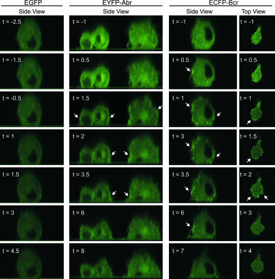
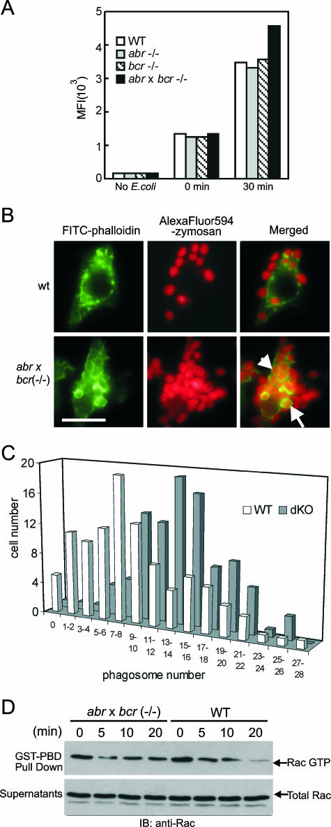
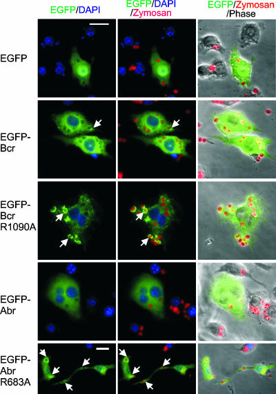
Small GTPases of the Rho family are key regulators of phagocytic leukocyte function. Abr and Bcr are homologous, multidomain proteins. Their C-terminal domain has GTPase-activating protein (GAP) activity that, in vitro, is specific for Rac and Cdc42. To address the in vivo relevance of these entire proteins, of which little is known, the current study examined the effect of the genetic ablation of Abr and Bcr in murine macrophages. The concomitant loss of Abr and Bcr induced multiple alterations of macrophage cellular behavior known to be under the control of Rac. Macrophages lacking both Abr and Bcr exhibited an atypical, elongated morphology that was reproduced by the ectopic expression of GAP domain mutant Abr and Bcr in a macrophage cell line and of constitutively active Rac in primary macrophages. A robust increase in colony-stimulating factor 1 (CSF-1)-directed motility was observed in macrophages deficient for both proteins and, in response to CSF-1 stimulation, Abr and Bcr transiently translocated to the plasma membrane. Phagocytosis of opsonized particles was also increased in macrophages lacking both proteins and correlated with sustained Rac activation. Bcr and Abr GAP mutant proteins localized around phagosomes and induced distinct phagocytic cup formation. These results identify Abr and Bcr as the only GAPs to date that specifically negatively regulate Rac function in vivo in primary macrophages.


Figures

FIG. 1.
FIG. 1.
Abr and Bcr reduce levels of activated Rac but not activated Cdc42. (A) Plasmids encoding Bcr and Abr as EGFP fusion proteins as well as EGFP alone were coexpressed with HA-tagged Rac1 in CHO cells. HA-Rac1 GTP was affinity precipitated with GST-Pak RBD. Total Rac levels and Abr and Bcr expression were analyzed from the supernatants after GST-Pak RBD precipitation. Top panel, HA-Rac1 GTP; middle panel, total HA-Rac1 protein; bottom panel, expression of EGF-Bcr, EGFP-Abr, and EGFP. One representative image of three independent experiments is shown. (B) The ratio of HA-Rac1 GTP levels to total HA-Rac1 levels was determined and then normalized to that of EGFP-expressing cells. Bars represent the mean ± standard deviation of at least three independent experiments. (C) EGFP-Bcr, EGFP-Abr, and EGFP were coexpressed with HA-Cdc42, and the relative activation of HA-Cdc42 was determined. Bars represent the mean ± standard deviation of two independent experiments.
FIG. 2.
FIG. 2.
Abnormally elongated morphology of macrophages lacking both Abr and Bcr is phenocopied by activated Rac. PEMs of the indicated genotypes were plated on glass chamber slides and incubated for 24 h in the absence of CSF-1 (A), or BMMs of the wild type and double null mutants were cultured on glass chamber slides for 1.5 h (B, upper panel) or 24 h (B, bottom panels) in the presence of 120 ng/ml CSF-1. F-actin was stained with FITC-phalloidin (green), and nuclei were stained with DAPI (blue in panel A, red in panel B). Note that the double null BMMs already started showing the elongated morphology after 1.5 h in culture. Bars, 20 μm (A and C), 25 μm (B, top and bottom panels), or 50 μm (B, middle panel). (C) Wild-type BMMs were electroporated with the indicated constructs. Arrows point to cells expressing V12Cdc42. Cells were stained for nuclei (blue) and F-actin (red); EGFP-expressing cells appear green.
FIG. 3.
FIG. 3.
GAP activity of Bcr or Abr is inhibited by mutation of conserved arginine and asparagines residues. GST fusion proteins tested include the wild-type Bcr and Abr GAP domains (bars labeled Bcr and Abr), Bcr R1090A (Bcr R/A), Abr R683A (Abr R/A), Bcr R1090A/N1202A (Bcr RN/AA), and Abr R683A/N795A (Abr RN/AA). Purified Bcr or Abr GAP domains were tested for Rac1 GAP activity. Results are expressed as the change with respect to Rac GTPase alone. Each GAP assay was performed in duplicate, and bars represent the mean ± standard deviation of two independent experiments.
FIG. 4.
FIG. 4.
GAP-defective mutants of Abr and Bcr induce abnormally elongated morphology in a macrophage-derived cell line, RAW 264.7. Cells were transfected with the indicated plasmids. After 24 h, cells were fixed, stained for nuclei, and analyzed with a fluorescence microscope. (A) Cells were visualized for EGFP (green) and nuclei (blue). Bars, 20 μm. EGFP-Abr R683A-expressing cells are shown at a higher magnification. (B) EGFP images (green) of cells were superimposed on those of phase-contrast images (black and white) to illustrate overall cell shape. Note that some of the cells expressing GAP mutants of Abr and Bcr (arrowheads) developed a highly elongated shape or multiple protrusions, while untransfected cells (arrows) remained round.
FIG. 5.
FIG. 5.
(abr × bcr)−/− macrophages have increased motility both in vitro and in vivo. (A) BMMs were added to the upper chamber of Transwells and allowed to migrate for 3 h. Random motility was measured in the presence of CSF-1 in both the upper and lower chambers of Transwells (white bars), and directed migration was measured in the presence of CSF-1 in the lower chamber (gray bars). Black bars represent motility in the absence of CSF-1 in both the upper and lower chambers. Values shown are the mean ± standard deviation (n = 2 per genotype), and each experiment was performed in duplicate. The differences in CSF-1-directed migration between the wild-type and abr/ BMMs and between the wild-type and (abr × bcr)−/− BMMs were statistically significant (P < 0.05; two-tailed Student t test, Bonferroni adjustment). (B) The number of macrophages in peritoneal exudates from wild-type or double null mutant mice (n = 3) was compared 4 days after intraperitoneal thioglycolate injection and was greater in double null mutant mice (P < 0.05).
FIG. 6.
FIG. 6.
Abr and Bcr translocate to the plasma membrane upon stimulation with CSF-1. Wild-type BMMs were transduced with lentivirus encoding the indicated proteins. The transduced BMMs were starved of CSF-1 overnight and allowed to attach to the fibronectin-coated surface of a glass-bottom petri dish. Serial confocal images in the x-z mode (side view) and x-y mode (top view), before and after the addition of CSF-1 to the cells, were collected every 30 seconds. Each number represents a time point in minutes before (designated with a minus sign) and after (designated with a plus sign) CSF-1 addition. CSF-1 was added between t = −1 and t = 1 for each series of images. The nucleus of each cell was identified by the dark area in the center of the cell, which lacks expression of the fusion proteins. The plane of focus for images captured in the x-z mode was through the nucleus of the cell, while that of the images captured in the x-y mode was near the attachment of the cell to the glass. The arrows mark areas of fluorescence intensification that occurred at the periphery of the cells.
FIG. 7.
FIG. 7.
Combined loss of Abr and Bcr results in increased phagocytosis by macrophages and is associated with sustained Rac activation. (A) BMMs were incubated with FITC-E. coli at a ratio of 1:25 in serum-free medium and analyzed by FACS. MFIs of FITC fluorescence from FACS were plotted on the y axis. (B) Representative images of wild-type and double null mutant cells phagocytosing opsonized zymosan particles. F-actin was stained with FITC-phalloidin (green), and zymosan was coupled with AlexaFluor (red). Red and green images are merged to identify the internalized zymosan particles. The external particles appear bright red, whereas the internalized zymosan particles appear orange and are surrounded by F-actin-rich phagocytic cups (arrows point to examples). Bar, 20 μm. (C) Number of phagosomes containing opsonized zymosan. The amount of internalized particles in each cell was counted and plotted against the number of cells containing that amount of particles. The difference in phagosome number distribution between wild-type and the double null BMMs was statistically significant (P < 0.01). (D) BMMs of the indicated genotypes were preincubated with opsonized FITC-E. coli, allowed to phagocytose for the indicated time, and assayed for activated and total Rac as described for Fig. 1.
FIG. 8.
FIG. 8.
GAP-defective mutant Bcr and Abr localize to phagocytic cups during phagocytosis of opsonized zymosan particles. After transfection with the indicated plasmids, RAW cells were serum starved and then incubated with Texas Red-coupled zymosan particles at a ratio of 1:25. EGFP-expressing cells appear green, zymosan is red, and nuclei are blue. Images with different fluorescences were merged, as indicated above the panels, to identify EGFP signal localizing to phagosomes (arrowheads point to examples). Bars, 20 μm. Note that the images of the EGFP-Abr R683A-transfected cells are shown at a lower magnification than the others, in order to capture one elongated cell.

Similar articles

Cited by

References

    1. Aghazadeh, B., K. Zhu, T. J. Kubiseski, G. A. Liu, T. Pawson, Y. Zheng, and M. K. Rosen. 1998. Structure and mutagenesis of the Dbl homology domain. Nat. Struct. Biol. 5:1098-1107. - PubMed
    1. Allen, W. E., G. E. Jones, J. W. Pollard, and A. J. Ridley. 1997. Rho, Rac and Cdc42 regulate actin organization and cell adhesion in macrophages. J. Cell Sci. 110:707-720. - PubMed
    1. Allen, W. E., D. Zicha, A. J. Ridley, and G. E. Jones. 1998. A role for Cdc42 in macrophage chemotaxis. J. Cell Biol. 141:1147-1157. - PMC - PubMed
    1. Benard, V., and G. M. Bokoch. 2002. Assay of Cdc42, Rac, and Rho GTPase activation by affinity methods. Methods Enzymol. 345:349-359. - PubMed
    1. Bernards, A. 2003. GAPs galore! A survey of putative Ras superfamily GTPase activating proteins in man and Drosophila. Biochim. Biophys. Acta 1603:47-82. - PubMed

Publication types

MeSH terms