. 2007 Feb;27(3):854-63.
doi: 10.1128/MCB.01400-06. Epub 2006 Nov 20.

Intramolecular regulation of phospholipase C-gamma1 by its C-terminal Src homology 2 domain

Affiliations

Intramolecular regulation of phospholipase C-gamma1 by its C-terminal Src homology 2 domain

Karen DeBell et al. Mol Cell Biol. 2007 Feb.

Abstract

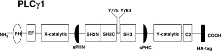
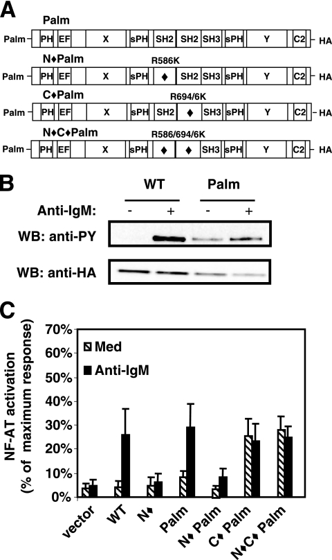
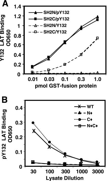
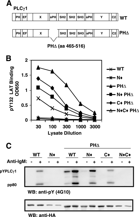
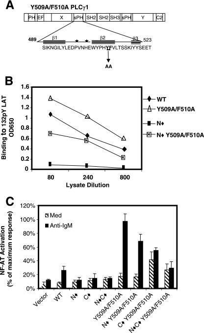
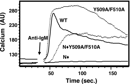
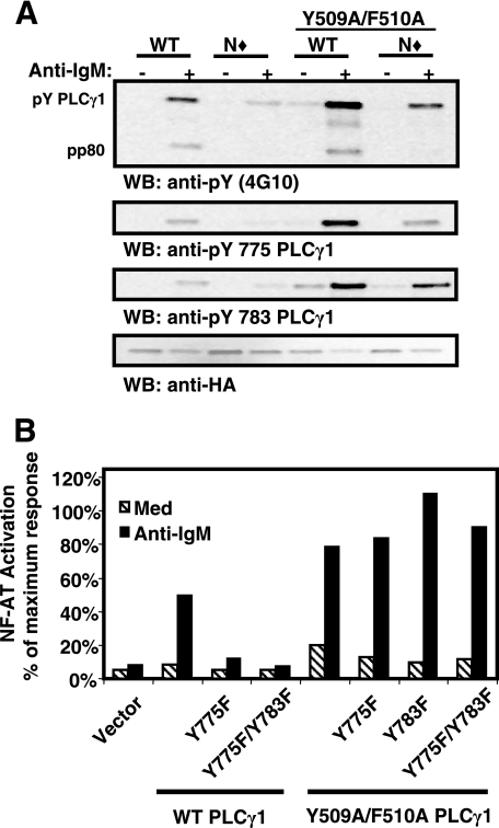
Phosphoinositide-specific phospholipase C-gamma1 (PLC-gamma1) is a key enzyme that governs cellular functions such as gene transcription, secretion, proliferation, motility, and development. Here, we show that PLC-gamma1 is regulated via a novel autoinhibitory mechanism involving its carboxy-terminal Src homology (SH2C) domain. Mutation of the SH2C domain tyrosine binding site led to constitutive PLC-gamma1 activation. The amino-terminal split pleckstrin homology (sPHN) domain was found to regulate the accessibility of the SH2C domain. PLC-gamma1 constructs with mutations in tyrosine 509 and phenylalanine 510 in the sPHN domain no longer required an intact amino-terminal Src homology (SH2N) domain or phosphorylation of tyrosine 775 or 783 for activation. These data are consistent with a model in which the SH2C domain is blocked by an intramolecular interaction(s) that is released upon cellular activation by occupancy of the SH2N domain.


Figures

FIG. 1.
FIG. 1.
Schematic representation of PLC-γ1 structure. EF, EF hand domain.
FIG. 2.
FIG. 2.
Raft-targeted PLC-γ1 is constitutively active when the SH2C domain is mutated. (A) Representation of raft-targeted PLC-γ1 constructs bearing an N-terminal palmitoylation signal sequence (Palm PLC-γ1). Mutated domains are indicated by a diamond (⧫). The amino acid substitution(s) used to inactivate each SH2 domain is shown for each construct. (B) P10-14 cells transiently transfected with WT or Palm PLC-γ1 were treated with medium or anti-IgM. Lysates were immunoprecipitated with anti-HA antibody, Western blotted for pan-tyrosine phosphorylation with the 4G10 antibody, and reprobed with anti-HA antibody. PY, phosphotyrosine. (C) P10-14 cells were transiently cotransfected with the indicated PLC-γ1 construct and an NFAT luciferase reporter and stimulated with medium or anti-IgM as indicated. The measured luciferase activity was normalized to the activity obtained for treatment with phorbol myristate acetate plus ionomycin. The data presented are the means and standard errors of the means for three separate experiments, with each condition assayed in duplicate. WB, Western blot; Med, medium.
FIG. 3.
FIG. 3.
The structure of PLC-γ1 regulates the ability of the SH2C domain to interact with proteins. (A) Serially diluted GST-SH2N and GST-SH2C fusion proteins were placed in wells of a streptavidin plate that had been coated with either biotinylated nonphosphorylated Y132 LAT (Y132) or phosphorylated pY132 LAT (pY132) peptide as indicated. Binding was detected with anti-GST rabbit serum and HRP-conjugated second antibody. The data are representative of three separate experiments with samples tested in duplicate. (B) Serially diluted lysates from P10-14 cells stably transfected with the indicated PLC-γ1 constructs were assayed for their ability to bind to the pY132 LAT peptide as described for panel A, except that binding was detected with an anti-HA antibody. The data are presented as the average optical densities at 650 nm (OD650) for two replicate wells in a single experiment and are representative of at least three separate experiments.
FIG. 4.
FIG. 4.
The amino-terminal split pleckstrin homology domain regulates the binding activity of the Src homology domain region of PLC-γ1. (A) Schematic diagram depicting the deletion of the amino-terminal split pleckstrin homology domain in PLC-γ1. (B) Lysates from P10-14 cells stably expressing PLC-γ1 PHΔ in combination with mutations of SH2N and/or SH2C were tested by ELISA for binding to pY132 LAT as described for Fig. 3B. (C) P10-14 cells stably expressing the indicated PLC-γ1 constructs were stimulated with medium or anti-IgM, lysed, and treated as described for Fig. 2B. EF, EF hand domain; aa, amino acids; OD650, optical density at 650 nm; WB, Western blot.
FIG. 5.
FIG. 5.
Amino acids Y509 and F510 in the sPHN domain participate in regulating SH2C domain availability. (A) Schematic diagram of PLC-γ1 displaying an expanded sPHN domain. Specifically noted are two amino acid residues, Y509 and F510 in the second β-sheet, that were mutated to alanine. Stars mark potential PI(4,5)P2 binding sites (18). (B) Serially diluted lysates from P10-14 cells transiently transfected with PLC-γ1 constructs bearing the indicated mutations in the sPHN domain were tested for binding to pY132 LAT by ELISA as described for Fig. 3B. (C) P10-14 cells were transiently transfected with the indicated PLC-γ1 construct and an NFAT luciferase reporter construct and then assayed for their response to treatment with anti-IgM as described for Fig. 2C. OD650, optical density at 650 nm; Med, medium.
FIG. 6.
FIG. 6.
Mutation of Y509 and F510 alters the BCR-induced calcium response. P10-14 cells stably expressing the indicated PLC-γ1 constructs were loaded with Indo-1/AM and stimulated with anti-IgM. The ratio of fluorescence emission at 405 nm to that at 495 nm is plotted over a 3-minute time period and is shown as arbitrary calcium units (AU).
FIG. 7.
FIG. 7.
Mutation of Y509 and F510 alleviates the requirement for Y775 and Y783 phosphorylation in PLC-γ1 activation. (A) Stable P10-14 transfectants were treated with medium or anti-IgM, lysed, and immunoprecipitated with anti-HA antibody. Samples were divided for resolution on individual sodium dodecyl sulfate-polyacrylamide gel electrophoresis gels and immunoblotted with antiphosphotyrosine (4G10), anti-pY775 PLC-γ1, anti-pY783 PLC-γ1, and anti-HA antibodies. (B) P10-14 cells were transiently transfected with the indicated PLC-γ1 constructs plus an NFAT luciferase reporter construct and then assayed for their response to treatment with medium or anti-IgM antibody as described for Fig. 2C. The data are representative of at least three independent experiments. WB, Western blot; Med, medium.
FIG. 8.
FIG. 8.
Intramolecular interactions regulate PLC-γ1 catalytic activity in unstimulated lymphocytes. (A) In unstimulated cells, PLC-γ1 is cytoplasmic. The carboxy-terminal SH2 (2C) domain and the sPHN domain cooperate to maintain PLC-γ1 in an inactive configuration. Intramolecular interactions between the SH2C domain and PLC-γ1 may position the PCI sequence (•) in proximity to the catalytic domain(s), inhibiting function. SH2C intramolecular binding also limits the ability of the SH2C domain to interact with phosphorylated target proteins. (B) Upon antigen receptor engagement, the amino-terminal SH2 (2N) domain interacts with its target protein (phospho-LAT in T cells or phospho-BLNK in B cells). This interaction is required to induce the release of the SH2C domain from its intramolecular interaction and free the catalytic domain(s) from inhibition by the PCI sequence. The SH2C domain is now able to interact with target proteins such as Slp76 and c-Cbl, and PLC-γ1 is phosphorylated on Y775 and Y783 (formula image
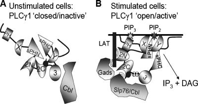
). Once released from intramolecular inhibition, the catalytic domains act in concert to hydrolyze PI(4,5)P2 (PIP2).

References

    1. Braiman, A., M. Barda-Saad, C. L. Sommers, and L. E. Samelson. 2006. Recruitment and activation of PLCgamma1 in T cells: a new insight into old domains. EMBO J. 25:774-784. - PMC - PubMed
    1. Carpenter, G., and Q. Ji. 1999. Phospholipase C-gamma as a signal-transducing element. Exp. Cell Res. 253:15-24. - PubMed
    1. Chang, J. S., H. Seok, T. K. Kwon, D. S. Min, B. H. Ahn, Y. H. Lee, J. W. Suh, J. W. Kim, S. Iwashita, A. Omori, S. Ichinose, O. Numata, J. K. Seo, Y. S. Oh, and P. G. Suh. 2002. Interaction of elongation factor-1alpha and pleckstrin homology domain of phospholipase C-gamma 1 with activating its activity. J. Biol. Chem. 277:19697-19702. - PubMed
    1. Choi, J. H., W. P. Hong, S. Yun, H. S. Kim, J. R. Lee, J. B. Park, Y. S. Bae, S. H. Ryu, and P. G. Suh. 2005. Grb2 negatively regulates epidermal growth factor-induced phospholipase C-gamma1 activity through the direct interaction with tyrosine-phosphorylated phospholipase C-gamma1. Cell. Signal. 17:1289-1299. - PubMed
    1. Crabtree, G. R., and N. A. Clipstone. 1994. Signal transmission between the plasma membrane and nucleus of T lymphocytes. Annu. Rev. Biochem. 63:1045-1083. - PubMed

MeSH terms

LinkOut - more resources