. 2006 Dec 5;103(49):18450-7.
doi: 10.1073/pnas.0605697103. Epub 2006 Nov 20.

The root endophytic fungus Piriformospora indica requires host cell death for proliferation during mutualistic symbiosis with barley

Affiliations

The root endophytic fungus Piriformospora indica requires host cell death for proliferation during mutualistic symbiosis with barley

Sachin Deshmukh et al. Proc Natl Acad Sci U S A. .

Abstract

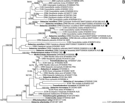
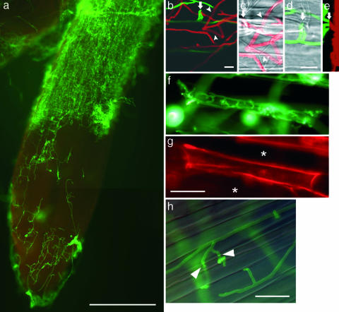
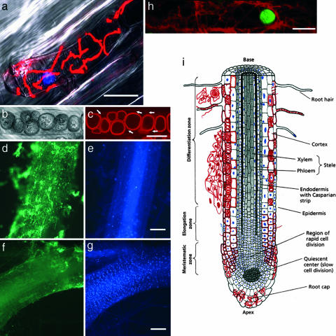
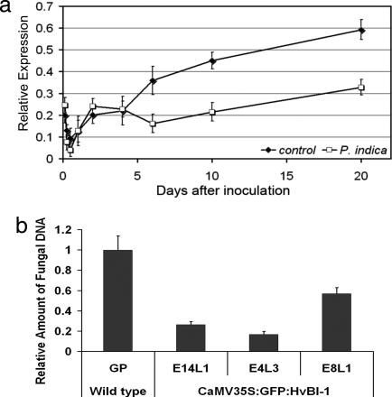
Fungi of the recently defined order Sebacinales (Basidiomycota) are involved in a wide spectrum of mutualistic symbioses (including mycorrhizae) with various plants, thereby exhibiting a unique potential for biocontrol strategies. The axenically cultivable root endophyte Piriformospora indica is a model organism of this fungal order. It is able to increase biomass and grain yield of crop plants. In barley, the endophyte induces local and systemic resistance to fungal diseases and to abiotic stress. To elucidate the lifestyle of P. indica, we analyzed its symbiotic interaction and endophytic development in barley roots. We found that fungal colonization increases with root tissue maturation. The root tip meristem showed no colonization, and the elongation zone showed mainly intercellular colonization. In contrast, the differentiation zone was heavily infested by inter- and intracellular hyphae and intracellular chlamydospores. The majority of hyphae were present in dead rhizodermal and cortical cells that became completely filled with chlamydospores. In some cases, hyphae penetrated cells and built a meshwork around plasmolyzed protoplasts, suggesting that the fungus either actively kills cells or senses cells undergoing endogenous programmed cell death. Seven days after inoculation, expression of barley BAX inhibitor-1 (HvBI-1), a gene capable of inhibiting plant cell death, was attenuated. Consistently, fungal proliferation was strongly inhibited in transgenic barley overexpressing GFP-tagged HvBI-1, which shows that P. indica requires host cell death for proliferation in differentiated barley roots. We suggest that the endophyte interferes with the host cell death program to form a mutualistic interaction with plants.


Conflict of interest statement

The authors declare no conflict of interest.

Figures

Fig. 1.
Fig. 1.
Phylogenetic placement of the strains tested in this study within the Sebacinales, estimated by maximum likelihood from an alignment of nuclear rDNA coding for the 5′ terminal domain of the ribosomal large subunit. Branch support is given by nonparametric maximum likelihood bootstrap (first numbers) and by posterior probabilities estimated by Bayesian Markov chain Monte Carlo (second numbers). Support values of <50% are omitted or indicated by an asterisk. The tree was rooted according to the results of ref. , and subgroups discussed in ref. are denoted with “A” and “B.” Sequences of the strains used in this study are indicated by black circles. Sequences from morphologically determined specimens or cultures are printed in bold. Sebacinalean sequences obtained from mycorrhizal plant roots are assigned to mycorrhizal types by the following acronyms: CVM, cavendishioid mycorrhiza (14); ECM, ectomycorrhiza; ERM, ericoid mycorrhiza; JMM, jungermannioid mycorrhiza; and ORM, orchid mycorrhiza. Proveniences are given as follows: A, Austria; AUS, Australia; CAN, Canada; CHN, People's Republic of China; ECU, Ecuador; EST, Estonia; FRA, France; GER, Germany; GUY, Guyana; IND, India; MEX, Mexico; NOR, Norway; and SPA, Spain.
Fig. 2.
Fig. 2.
Infestation pattern of P. indica in barley roots. (a) By 8 dai, hyphae excessively occupy rhizodermal and cortical cells of the differentiation zone. The elongation zone is less colonized, with occasional intercellular subepidermal hyphal structures. The root cap is heavily infested with hyphae. (be) After penetration (arrows) fungal hyphae colonize the subepidermal layer. (b) To better visualize the position of hyphae in the z axis, a confocal laser scanning image consisting of 30 frames of adjacent focal planes (z axis) was displayed as a maximum projection with the fluorescent signal of the wheat germ agglutinin-stained fungal hyphae displayed in red for the upper (abaxial) 15 frames and in green for the lower (adaxial, subepidermal) 15 frames. (c and d) For visualization of plant cell walls, two close-up bright-field images of two different focal planes are superimposed with the respective frames of the fluorescence images. Intercellular hyphae start branching and proliferate within the subepidermal space. (c) Subepidermal hyphae crossing cell walls (arrowheads) without exhibiting morphological changes (e.g., neck formation, as in h) revealing their periclinal localization. (d) The upper focal plane is characterized by hyphae penetrating the anticlinal space of adjacent rhizodermis cells. (e) Projection of the fluorescent signals of c and d in the y axis (vertical) and z axis (horizontal). Absence of fluorescent signals between adaxial (green) and abaxial hyphae (red) indicates a layer of rhizodermal cells free from hyphae. The penetration site is indicated by an arrow. (f) Colonization of a single cell within young differentiated tissue. After penetration, the cell is completely filled with intracellular hyphae before the colonization of adjacent cells. (g) The cell wall of the colonized cell is strongly stained with Congo red because of better dye accessibility compared with noncolonized neighbor cells (asterisks). Penetrated cells did not show autofluorescence. (h) Intracellular mycelium in mature root tissue. Overlay of bright-field image and fluorescence image. Intracellular hyphae form necks (arrowheads) at sites of cell wall crossing. Fungal structures are visualized by WGA-AF 488. [Scale bars: a, 300 μm; b and d, 10 μm (c and d are of the same scale); fh, 30 μm.]
Fig. 3.
Fig. 3.
Association of fungal structures with living and dead cells of the host tissue. (a) Fungal hyphae swathe a plant protoplast, which undergoes cytoplasmic shrinkage. Hyphae and nucleus stained with WGA-TMR and DAPI, respectively, are superimposed with the bright-field image. (b) Bright-field interference contrast image of chlamydospores in a root cortex cell. (c) Fluorescence image of the same cell stained with fuchsin-lactic acid. Arrows indicate hyphae on which the chlamydospores are formed. (dg) Root colonization spatially associated with the absence of intact plant nuclei. Root segments (60 hours after inoculation) double-stained for intact plant nuclei (DAPI; e and g) and fungal hyphae (WGA-AF 488; d and f). (d and e) A root segment heavily colonized by fungal hyphae (d) contains only a few DAPI-stained nuclei (e). (f and g) A root segment with minor fungal colonization (f) contains a high number of DAPI-stained nuclei (g). (h) Hyphae swathing a cortical cell protoplast with a TUNEL-positive (green) nucleus. (i) Schematic drawing of a P. indica-infested root showing the different tissues and the associated colonization pattern, with hyphae depicted in red and DAPI-positive plant nuclei depicted in blue. (Scale bars: a, 30 μm; c, 10 μm; dg, 300 μm; and h, 20 μm.) [Modified from ref. (Copyright 1998, Sinauer, Sunderland, MA).]
Fig. 4.
Fig. 4.
Influence of HvBI-1 on the development of P. indica in barley roots. (a) Quantitative PCR analysis of HvBI-1 expression. As compared with noncolonized roots, expression of the gene is significantly lower from 7 dai onward up to 20 dai. Error bars represent standard deviations. (b) The relative amount of P. indica DNA in transgenic GFP-HvBI-1 roots was determined at 20 dai. Error bars represent standard deviations. GP represents wild-type plants (Golden Promise). E14L1, E4L3, and E8L1 represent independent transgenic GFP-HvBI-1 GP lines, with five plants tested per line. All three lines were significantly different from the wild type (P <0.005, Student's t test). Similar results were obtained in three experiments with plants of an independent transgenic GFP-HvBI-1 line.

Similar articles

Cited by

References

    1. Kogel KH, Franken P, Hückelhoven R. Curr Opin Plant Biol. 2006;9:358–363. - PubMed
    1. Harrison MJ. Annu Rev Microbiol. 2005;59:19–42. - PubMed
    1. Borowicz VA. Ecology. 2001;82:3057–3068.
    1. Arnold AE, Mejia LC, Kyllo D, Rojas EI, Maynard Z, Robbins N, Herre EA. Proc Natl Acad Sci USA. 2003;100:15649–15654. - PMC - PubMed
    1. Müller CB, Krauss J. Curr Opin Plant Biol. 2005;8:450–456. - PubMed

LinkOut - more resources