

Muscle architecture and functional anatomy of the pelvic limb of the ostrich (Struthio camelus)
- PMID: 17118064
- PMCID: PMC2048997
- DOI: 10.1111/j.1469-7580.2006.00658.x
Muscle architecture and functional anatomy of the pelvic limb of the ostrich (Struthio camelus)
Abstract
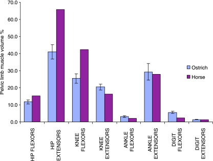
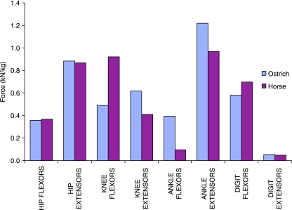
The functional anatomy of the pelvic limb of the ostrich (Struthio camelus) was investigated in order to assess musculoskeletal specialization related to locomotor performance. The pelvic limbs of ten ostriches were dissected and detailed measurements of all muscle tendon units of the pelvic limb were made, including muscle mass, muscle length, fascicle length, pennation angle, tendon mass and tendon length. From these measurements other muscle properties such as muscle volume, physiological cross-sectional area (PCSA), tendon cross-sectional area, maximum isometric muscle force and tendon stress were derived, using standard relationships and published muscle data. Larger muscles tended to be located more proximally and had longer fascicle lengths and lower pennation angles. This led to an expected proximal to distal reduction in total muscle mass. An exception to this trend was the gastrocnemius muscle, which was found to have the largest volume and PCSA and also had the highest capacity for both force and power production. Generally high-power muscles were located more proximally in the limb, while some small distal muscles (tibialis cranialis and flexor perforatus digiti III), with short fibres, were found to have very high force generation capacities. The greatest proportion of pelvic muscle volume was for the hip extensors, while the highest capacity for force generation was observed in the extensors of the ankle, many of which were also in series with long tendons and thus were functionally suited to elastic energy storage.
Figures
Similar articles
-
Ontogenetic scaling of pelvic limb muscles, tendons and locomotor economy in the ostrich (Struthio camelus).J Exp Biol. 2019 Sep 3;222(Pt 17):jeb182741. doi: 10.1242/jeb.182741. J Exp Biol. 2019. PMID: 31350301 Free PMC article.
-
Functional specialisation of pelvic limb anatomy in horses (Equus caballus).J Anat. 2005 Jun;206(6):557-74. doi: 10.1111/j.1469-7580.2005.00420.x. J Anat. 2005. PMID: 15960766 Free PMC article.
-
Anatomical description of the muscles of the pelvic limb in the ostrich (Struthio camelus).Anat Histol Embryol. 2004 Apr;33(2):100-14. doi: 10.1111/j.1439-0264.2003.00522.x. Anat Histol Embryol. 2004. PMID: 15027951
-
Review of the methods used for calculating physiological cross-sectional area (PCSA) for ecological questions.J Morphol. 2020 Jul;281(7):778-789. doi: 10.1002/jmor.21139. Epub 2020 May 6. J Morphol. 2020. PMID: 32374505 Review.
-
Functional and architectural complexity within and between muscles: regional variation and intermuscular force transmission.Philos Trans R Soc Lond B Biol Sci. 2011 May 27;366(1570):1477-87. doi: 10.1098/rstb.2010.0359. Philos Trans R Soc Lond B Biol Sci. 2011. PMID: 21502119 Free PMC article. Review.
Cited by
-
Functional specialization and ontogenetic scaling of limb anatomy in Alligator mississippiensis.J Anat. 2010 Apr;216(4):423-45. doi: 10.1111/j.1469-7580.2009.01202.x. Epub 2010 Feb 10. J Anat. 2010. PMID: 20148991 Free PMC article.
-
How to walk carrying a huge egg? Trade-offs between locomotion and reproduction explain the special pelvis and leg anatomy in kiwi (Aves; Apteryx spp.).J Anat. 2019 Dec;235(6):1045-1056. doi: 10.1111/joa.13072. Epub 2019 Aug 20. J Anat. 2019. PMID: 31432515 Free PMC article.
-
Adaptations for economical bipedal running: the effect of limb structure on three-dimensional joint mechanics.J R Soc Interface. 2011 May 6;8(58):740-55. doi: 10.1098/rsif.2010.0466. Epub 2010 Oct 28. J R Soc Interface. 2011. PMID: 21030429 Free PMC article.
-
Limb Kinematics, Kinetics and Muscle Dynamics During the Sit-to-Stand Transition in Greyhounds.Front Bioeng Biotechnol. 2018 Nov 16;6:162. doi: 10.3389/fbioe.2018.00162. eCollection 2018. Front Bioeng Biotechnol. 2018. PMID: 30505834 Free PMC article.
-
On the 3D Nature of the Magpie (Aves: Pica pica) Functional Hindlimb Anatomy During the Take-Off Jump.Front Bioeng Biotechnol. 2021 Jun 29;9:676894. doi: 10.3389/fbioe.2021.676894. eCollection 2021. Front Bioeng Biotechnol. 2021. PMID: 34268296 Free PMC article.
References
-
- Alexander RM, Maloiy GMO, Njau R, Jayes AS. Mechanics of running in the ostrich (Struthio camelus) J Zool Lond. 1979;187:169–178.
-
- Alexander RM. Energy-saving mechanisms in walking and running. J Exp Biol. 1991;160:55–69. - PubMed
-
- Alexander RM. Tendon elasticity and muscle function. Comp Biochem Physiol – Part A: Mol Integrative Physiol. 2002;133:1001–1011. - PubMed
-
- Askew GN, Marsh RL. The mechanical power output of the pectoralis muscle of blue-breasted quail (Coturnix chinensis): the in vivo length cycle and its implications for muscle performance. J Exp Biol. 2001;204:3587–3600. - PubMed
-
- Bennett MB, Ker RF, Dimery NJ, Alexander RM. Mechanical properties of various mammalian tendons. J Zool Lond. 1986;209:537–548.
Publication types
MeSH terms
LinkOut - more resources
Full Text Sources
