Review
. 2006;4 Suppl 1(Suppl 1):S6.
doi: 10.1186/1477-7827-4-S1-S6.

A critical period of progesterone withdrawal precedes menstruation in macaques

Affiliations
Review

A critical period of progesterone withdrawal precedes menstruation in macaques

Ov D Slayden et al. Reprod Biol Endocrinol. 2006.

Abstract

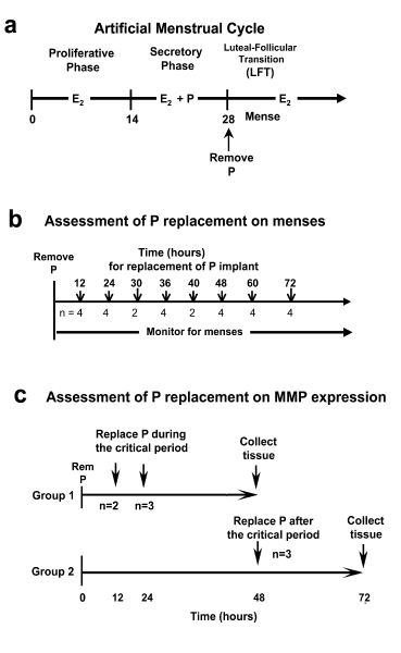
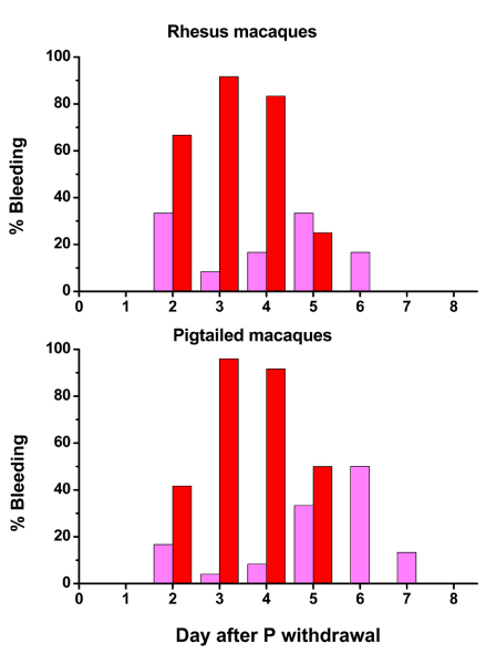
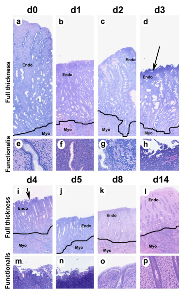
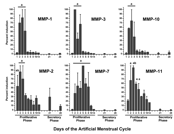
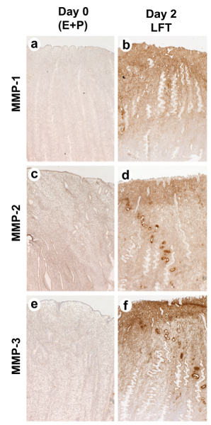
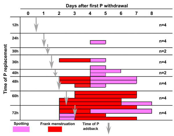
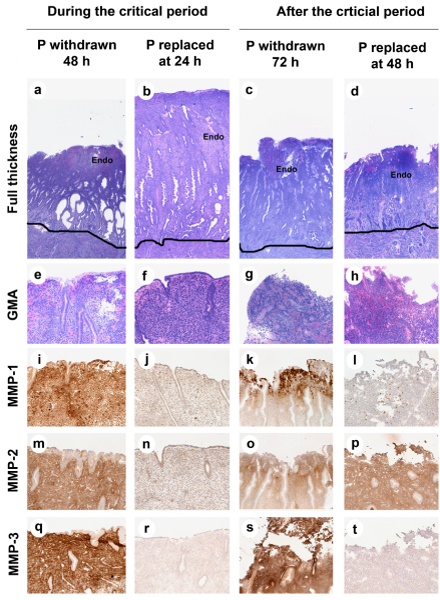
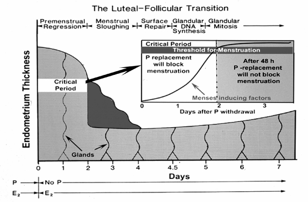
Macaques are menstruating nonhuman primates that provide important animal models for studies of hormonal regulation in the uterus. In women and macaques the decline of progesterone (P) at the end of the cycle triggers endometrial expression of a variety of matrix metalloproteinase (MMP) enzymes that participate in tissue breakdown and menstrual sloughing. To determine the minimal duration of P withdrawal required to induce menses, we assessed the effects of adding P back at various time points after P withdrawal on both frank bleeding patterns and endometrial MMP expression. Artificial menstrual cycles were induced by treating the animals sequentially with implants releasing estradiol (E2) and progesterone (P). To assess bleeding patterns, P implants were removed at the end of a cycle and then added back at 12, 24, 30, 36, 40, 48, 60, or 72 hours (h) after the initial P withdrawal. Observational analysis of frank bleeding patterns showed that P replacement at 12 and 24 h blocked menses, replacement at 36 h reduced menses but replacement after 36 h failed to block menses. These data indicate that in macaques, a critical period of P withdrawal exists and lasts approximately 36 h. In other similarly cycled animals, we withdrew P and then added P back either during (12-24 h) or after (48 h) the critical period, removed the uterus 24 h after P add back and evaluated endometrial MMP expression. Immunocytochemistry showed that replacement of P during the critical period suppressed MMP-1, -2 and -3 expression along with menses, but replacement of P at 48 h, which failed to suppress mense, suppressed MMP-1 and MMP-3 but did not block MMP-2. We concluded that upregulation of MMPs is essential to menses induction, but that after the critical period, menses will occur even if some MMPs are experimentally blocked.


Figures

Figure 1
Figure 1
(A) Experimental protocols. The artificial menstrual cycle. Sequential treatment with E2 alone (Proliferative Phase) and then E2 + P (secretory phase) primes the endometrium for menstruation during the Luteal Follicular Transition (LFT) after P implant removal. (B) Assessment of P add back on menses. P implants were removed at the end of the cycle. P implants were then replaced in the animals at 12, 24, 30 36, 40, 48, 60, or 72 hours (h) after initial P withdrawal. Control animals (n = 1/time point) received blank Silastic implants. (C) Assessment of P add back on MMPs. P was withdrawn as described above, and then either replaced at 12 and 24 hours during the critical period (Group 1), or at 48 hours after the critical period (Group 2). Tissues were collected at 48 h for group 1 and 72 hours for group 2. Control animals did not have P implants replaces and tissues were collected at either 48 or 72 h after P withdrawal.
Figure 2
Figure 2
A rhesus macaque uterus from an early menstruating animal 48 hours after P withdrawal. For this photograph the uterus has been separated from the cervix and cut in half along the longitudinal axis. A black line has been drawn to delineate the endometrial-myometrial border. The beginning of tissue bleeding is evident in the functionalis zone. The black dashed line shows the full-thickness plane for histological sectioning. Endo = endometrium; Myo = myometrium.
Figure 3
Figure 3
Effects of P withdrawal on menstrual bleeding in rhesus and pigtailed macaques. In this chart, each bar represents the percentage of animals (n = 12) that were bleeding on each day of the LFT. Pink bars depicts swab-positive spotting, and red indicates frank menses.
Figure 4
Figure 4
Endometrial histology in the pigtailed macaque. Photomicrographs of GMA sections showing endometrial histology from pigtailed macaques on day 0,1, 2, 3, 4, 5, 8, and 14 of the artificial menstrual cycle. a-d and i-l show full-thickness sections all at the same magnification (25× original magnification). A line has been drawn to delineate the endometrial-myometrial border. High power photographs of the upper functionalis zone are shown in e-h and m-p (200× original magnification). At the end of the cycle (a, e) the endometrium is thick and the glands are sacculated due to the effect of P. P withdrawal induced endometrial shrinkage (day 1) and the beginning of bleeding by day 2 (note tissue breakdown in (g). The sloughing menstrual surface is clearly evident on d3 and d4 (Arrows in d and i point out menstrual surface). By day 5 the luminal surface is recovered with epithelial cells. During the proliferative phase (d8 and d 14) the endometrium regrows with tubular glands that show abundant mitotic cells (p). Endo = endometrium; Myo = myometrium.
Figure 5
Figure 5
Expression of MMP transcripts during the menstrual cycle in rhesus macaques. MMP expression was assayed by Northern Hybridization and expressed as a percent of the maximum for the group. Values were analyzed previously [6,7] by ANOVA followed by Fisher's Protected LSD. In each case MMPs were maximal during the menstrual period at the beginning of the proliferative phase. Bars with similar subscripts were significantly different (P < 0.05) from those without subsubscripts.
Figure 6
Figure 6
Immunostaining for MMP-1, MMP-2 and MMP-3 in the pigtailed macaque endometrium. Samples were collected either on day 0 (a,c,d) or on day 2, 48 hours after P withdrawal (b,d,f). Note the strong gradient of MMP immunostaining with the strongest signal in the upper and mid functionalis zone. Magnifications (50× original magnification).
Figure 7
Figure 7
Effect of P replacement during the LFT. This chart shows menstrual bleeding on each day after P withdrawal during the luteal-follicular transition. P implants were replaced at 12, 24, 30, 36 40 48 60 and 72 hours during this period (vertical gray arrows). Incidence of vaginal spotting (pink) and frank menstruation (red) on each day are shown as bars across the x axis. Each bar represents one animal. Replacement of P at 12, 24 and 30 hours blocked frank menses. Replacement of P at 36 h blocked bleeding in all but one animal, at 48 h failed to block bleeding in three out of 4 animals, and P replacement later than 48 h after P withdrawal failed to block bleeding in all animals.
Figure 8
Figure 8
Effect of P replacement on endometrial histology and MMP ICC. The photomicrographs are arranged as a grid with each treatment in columns and histological endpoints in each row. a-h: GMA sections showing endometrial histology (a-d; Magnification 100× original magnification and e-h; functionalis zone, Original Magnification 25×). i-t: ICC for MMPs. Strong staining for MMP1, MMP2, and MMP-3 was observed at 48 and 72 h in control animals. Replacement of P during, but not after, the critical period blocked menstrual breakdown (compare b to d and f to h). Replacement of P at 24 hours blocked expression of all three MMPs (j, n, r), but P replacement at 48 hours blocked MMP-1 and MMP-3 but not MMP-2 (l,p,t).
Figure 9
Figure 9
The Luteal-Follicular Transition. This diagram illustrates the various phases that follow P withdrawal in the primate endometrium, beginning with Premenstrual Regression, Menstrual Sloughing, Suface Repair, etc. The critical period is shown as a 2 day period during which menses-inducing factors build up to a threshold of inevitability.

References

    1. Brenner RM, West NB, McClellan MC. Estrogen and progestin receptors in the reproductive tract of male and female primates. Biol Reprod. 1990;42:11–19. doi: 10.1095/biolreprod42.1.11. - DOI - PubMed
    1. Brenner RM, McClellan MC, West NB, Novy MJ, Haluska GJ, Sternfeld MD. Estrogen and progestin receptors in the macaque endometrium. In: Bulletti C, Gurpide E, editor. The Primate Endometrium. NY: New York Academy of Sciences; 1991. pp. 149–166. - PubMed
    1. Brenner RM, Slayden OD. Cyclic changes in the primate oviduct and endometrium. In: Knobil E, Neill JD, editor. The Physiology of Reproduction. New York: Raven Press; 1994. pp. 541–569.
    1. Bartelmez GW. Cyclic changes in the endometrium of the rhesus monkey (Macaca mulatta) Contrib Embryol. 1951;34:99–144.
    1. Bartelmez GW. The phases of the menstrual cycle and their interpretation in terms of the pregnancy cycle. Am J Obstet Gynecol. 1957;74:931–955. - PubMed

Publication types