. 2007 Jan 15;73(2):270-8.
doi: 10.1016/j.bcp.2006.10.004. Epub 2006 Oct 13.

Calmodulin potentiates G beta gamma activation of phospholipase C-beta3

Affiliations

Calmodulin potentiates G beta gamma activation of phospholipase C-beta3

Jennifer S McCullar et al. Biochem Pharmacol. .

Abstract

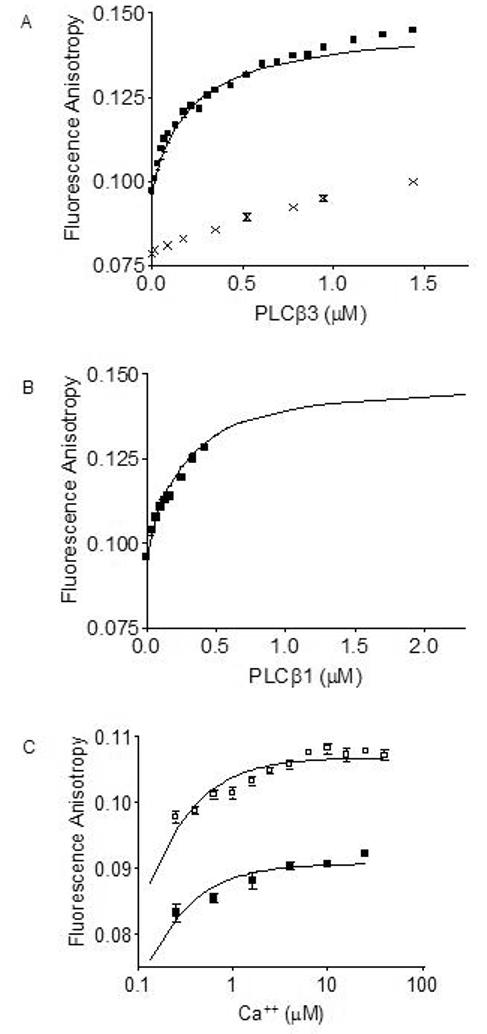
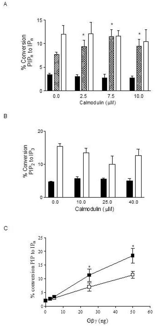
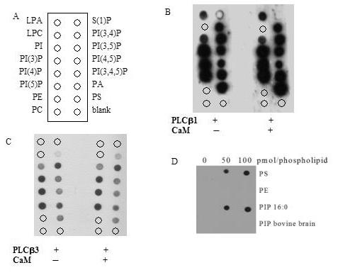
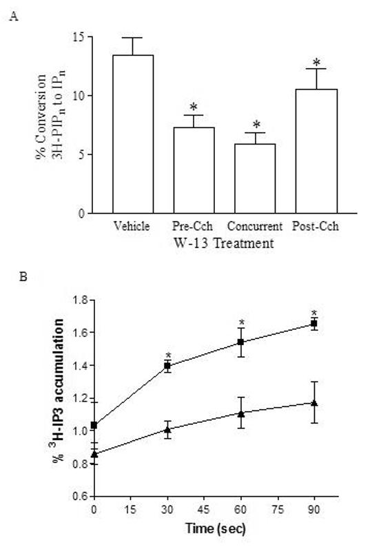
Phospholipase C-beta (PLC-beta) isozymes (EC 3.1.4.11) hydrolyze the membrane phospholipid phosphatidylinositol-4,5-bisphosphate to generate intracellular second messenger signaling molecules inositol-1,4,5-trisphosphate (IP3) and diacylglycerol (DAG) in response to receptor activation and other cellular stimuli. PLCbeta1 and PLCbeta3 isozymes were previously demonstrated to bind the calcium-sensitive molecule calmodulin [McCullar JS, Larsen SA, Millimaki RA, Filtz TM. Calmodulin is a phospholipase C-{beta} interacting protein. J Biol Chem 2003;278(36):33708-13]. We have now shown through fluorescence anisotropy that calmodulin/PLCbeta3 affinities increase with increasing calcium in a physiologically relevant concentration range. The bimolecular affinity constants for calmodulin interaction with PLCbeta1 or PLCbeta3 were estimated as 260 and 200 nM, respectively, from fluorescence anisotropy data. There was no effect of calmodulin on basal or G alpha q-stimulated catalytic activity for either isozyme. However, the interaction between calmodulin and PLCbeta3 leads to potentiation of activation by the G-protein beta gamma dimer in an in vitro assay. 1321N1 cells treated with calmodulin inhibitors concurrent with and post-stimulation of muscarinic receptors significantly reduced [3H]PIP hydrolysis. Together these data are suggestive of cooperative role for calmodulin in the G-protein beta gamma dimer-stimulated activity of PLCbeta3.


Figures

Figure 1
Figure 1
Binding of fluorescently-tagged calmodulin to PLCβ1 and PLCβ3 monitored by fluorescence anisotropy. Fluorescence anisotropy monitoring of 10 nM Alexa-calmodulin binding to increasing concentrations of (A) PLCβ3 (■) in the presence of 1 mM calcium, or presence of 1 mM EDTA (x). Lines were curvefit as described in Materials and Methods. Fluorescence anisotropy monitoring of 10 nM Alexa-calmodulin binding to increasing concentrations of (B) PLCβ1 (■) in the presence of 1 mM calcium. Lines were curvefit as described in Materials and Methods. KD values calculated are 260 nM for PLCβ1 interaction with calmodulin and 200 nM for PLCβ3 interaction with calmodulin. (C) Effect of increasing calcium concentrations on fluorescence anisotropy with 10 nM Alexa-calmodulin (■) or 10 nM Alexa-calmodulin plus 30 nM PLCβ3 (□). Fluorescence anisotropy (emission intensity) was measured at 525 nm with excitation at 490 nm at 20°C. Shown is representative data from two separate experiments. An apparent EC50 value of < 1 μM was calculated for calcium effects on PLCβ3/calmodulin interactions.
Figure 2
Figure 2
Effect of calmodulin on basal and G-protein-stimulated PLCβ activity in vitro. (A) Basal (solid bars), 50 ng (90 nM) Gβγ-stimulated- (hatched bars), and 30 ng (70 nM) GTPγSGαq-stimulated (white bars) PLCβ3 activity was quantitated in the presence of increasing concentrations of calmodulin. (B) Basal (solid bars) and 30 ng GTPγS-Gαq-stimulated (white bars) PLCβ1 activity was quantitated in the presence of increasing concentrations of calmodulin. Basal PLCβ activity was measured using PIP2 substrate in detergent/phospholipid vesicles. G-protein-stimulated PLCβ activity was measured in the presence of purified G protein subunits using PIP2 substrate in detergent/phospholipid vesicles. PLCβ activity was quantitated as percent conversion of [3H]PIP2 substrate to [3H]IPn (derivatives of inositol phosphate with the individual phosphate positions defined in accordance with the IUPAC convention) as described in Methods. (C) PLCβ3 activity was measured in the presence of increasing concentrations of Gβγ subunits with (■) or without (□) 10 μM calmodulin. Data shown are mean ± SEM of 3 to 5 separate experiments performed in triplicate. Asterisk (*) indicates treatment results significantly different from vehicle at p < 0.001.
Figure 3
Figure 3
Protein lipid overlay. (A) PIP strips with the following membrane lipids; lysophosphatidic acid, LPA, lysophosphatidylcholine, LPC, phosphaptidylinositol, PI, phosphatidylinositol-3-phosphate, PI(3)P, phosphatidylinositol-4-phosphate, PI(4)P, phosphatidylinositol-5-phosphate, PI(5)P, phosphatidylethanolamine, PE, phosphatidylcholine, PC, sphingo-1-phosphate, S(1)P, phosphatidylinositol-3,4-phosphate, PI(3,4)P, phosphatidylinositol-3,5-phosphate, PI(3,5)P, phosphatidylinositol-4,5-diphosphate, PI(4,5)P, phosphatidylinositol-3,4,5-trisphosphate, PI(3,4,5)P, phosphatidic acid, PA, phosphatidylserine, PS, and no lipid blank. PIP strips incubated with 0.5ug/ml of (B) PLCβ1 or (C) PLCβ3 in the absence or presence of 0.5ug/ml calmodulin. (D) Nitrocellulose membranes spotted with indicated amounts of the listed phosphospholipids (pmol) and incubated with 0.5 μg/ml PLCβ3. PE and PS have been isolated from bovine brain. Nitrocellulose membranes and PIP strips were washed, incubated with anti-PLCβ isoenzymes, and detected by Western blotting procedures as described in Materials and Methods. Data shown is representative of three similar experiments.
Figure 4
Figure 4
Calmodulin antagonist effects on carbachol-stimulated inositol phosphate accumulation under varying conditions of cell activation. (A) 1321N1 cells were stimulated for 20 minutes with 1 mM carbachol in the presence of 10 mM LiCl. Additionally, cells were either treated with dH20 (Vehicle) or treated with 100 μM W-13 for 20 minutes concurrent with carbachol addition (Concurrent), or treated for 10 minutes with 100 μM W-13 10 minutes after carbachol addition (Post-Cch). PLCβ activity was measured as percent of [3H]PIPn converted to IPn in whole cells, collected and quantitated as described in Materials and Methods. Shown is cumulative data from three experiments performed in triplicate. (B) 1321N1 cells were pre-treated with dH2O (Vehicle) or with 100 μM W-13 for 15 minutes. Cells were stimulated for 30, 60 and 90 seconds with 1 mM carbachol in the presence of 10 mM LiCl. PLCβ activity was measured as percent of [3H]PIPn converted to IP3 in whole cells, collected and quantitated as described in Materials and Methods. Shown is cumulative data from two experiments performed in triplicate. Asterisk (*) indicates W-13 pre-treatment results significantly different from vehicle pre-treatment at p < 0.001.

References

    1. McCullar JS, Larsen SA, Millimaki RA, Filtz TM. Calmodulin Is a Phospholipase C-{beta} Interacting Protein. J. Biol. Chem. 2003;278(36):33708–33713. - PubMed
    1. Rebecchi MJ, Pentyala SN. Structure, Function, and Control of Phosphoinositide-Specific Phospholipase C. Physiological Reviews. 2000;80:1291–1335. - PubMed
    1. Hwang JI, Oh YS, Shin KJ, Kim H, Ryu SH, Suh PG. Molecular cloning and characterization of a novel phospholipase C, PLC-eta. Biochem J. 2005;389(Pt 1):181–6. - PMC - PubMed
    1. Song C, Hu C-D, Masago M, Kariya K-i, Yamawaki-Kataoka Y, Shibatohge M, Wu D, Satoh T, Kataoka T. Regulation of a Novel Human Phospholipase C, PLCepsilon, through Membrane Targeting by Ras. J. Biol. Chem. 2001;276(4):2752–2757. - PubMed
    1. Saunders CM, Larman MG, Parrington J, Cox LJ, Royse J, Blayney LM, Swann K, Lai FA. PLC{zeta}: a sperm-specific trigger of Ca2+ oscillations in eggs and embryo development. Development. 2002;129(15):3533–3544. - PubMed

Publication types