. 2007 Feb 2;365(5):1393-406.
doi: 10.1016/j.jmb.2006.10.022. Epub 2006 Oct 13.

Structural transitions and thermodynamics of a glycine-dependent riboswitch from Vibrio cholerae

Affiliations

Structural transitions and thermodynamics of a glycine-dependent riboswitch from Vibrio cholerae

Jan Lipfert et al. J Mol Biol. .

Abstract

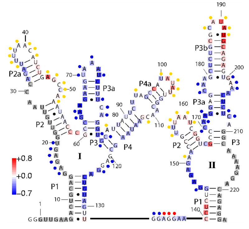
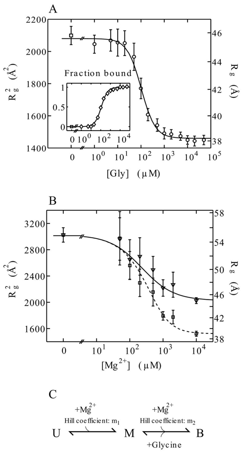
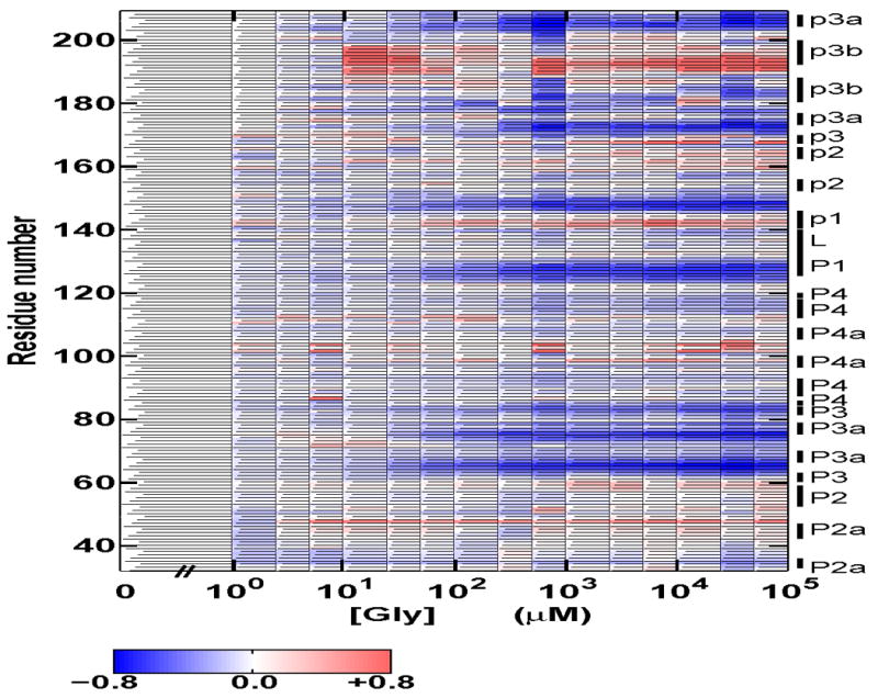
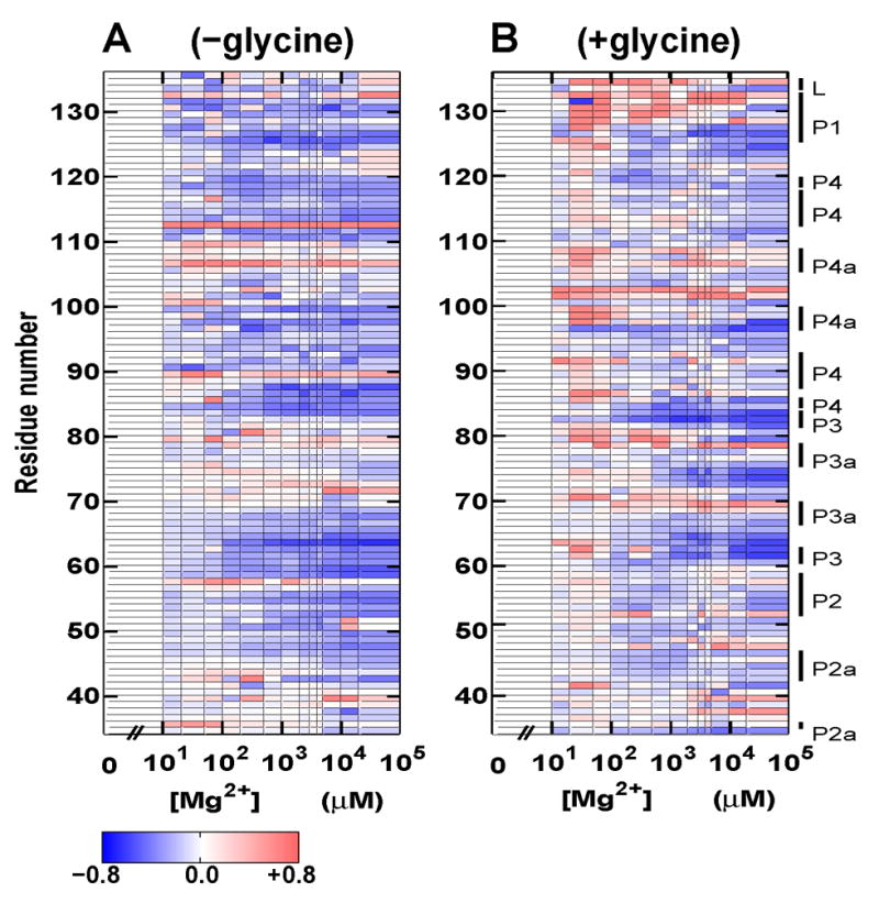
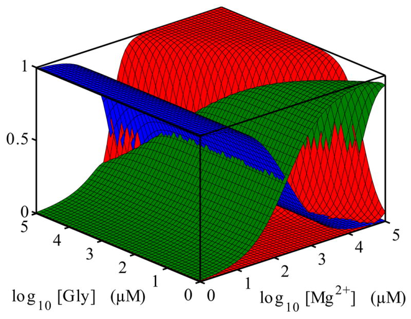
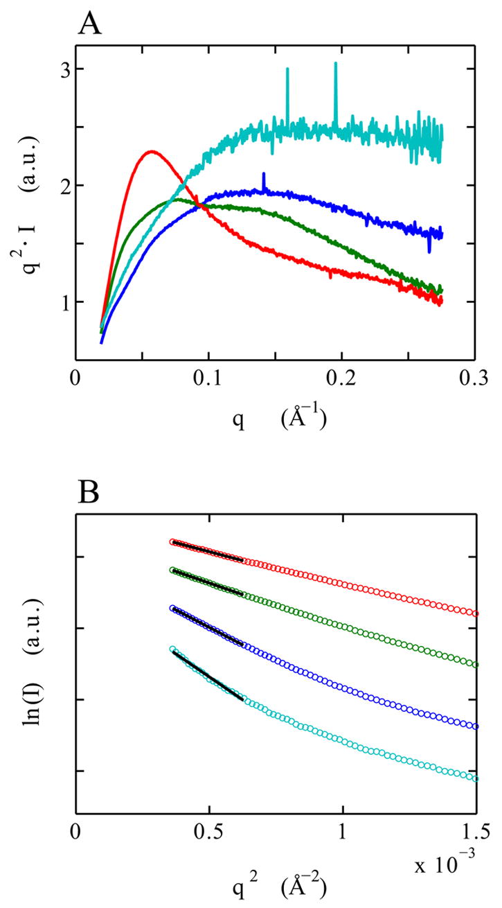
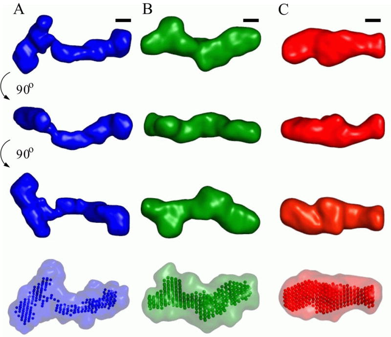
Riboswitches are complex folded RNA domains found in noncoding regions of mRNA that regulate gene expression upon small molecule binding. Recently, Breaker and coworkers reported a tandem aptamer riboswitch (VCI-II) that binds glycine cooperatively. Here, we use hydroxyl radical footprinting and small-angle X-ray scattering (SAXS) to study the conformations of this tandem aptamer as a function of Mg(2+) and glycine concentration. We fit a simple three-state thermodynamic model that describes the energetic coupling between magnesium-induced folding and glycine binding. Furthermore, we characterize the structural conformations of each of the three states: In low salt with no magnesium present, the VCI-II construct has an extended overall conformation, presumably representing unfolded structures. Addition of millimolar concentrations of Mg(2+) in the absence of glycine leads to a significant compaction and partial folding as judged by hydroxyl radical protections. In the presence of millimolar Mg(2+) concentrations, the tandem aptamer binds glycine cooperatively. The glycine binding transition involves a further compaction, additional tertiary packing interactions and further uptake of magnesium ions relative to the state in high Mg(2+) but no glycine. Employing density reconstruction algorithms, we obtain low resolution 3-D structures for all three states from the SAXS measurements. These data provide a first glimpse into the structural conformations of the VCI-II aptamer, establish rigorous constraints for further modeling, and provide a framework for future mechanistic studies.


Figures

Fig. 1
Fig. 1
Secondary structure of the VCI-II riboswitch tandem aptamer from Mandal, et. al. The color code mapped onto the secondary structure overlaying the letters represents relative solvent exposure measured by hydroxyl radical footprinting cleavage for the high glycine B state (average over data points at 10, 25, 50, and 100 mM glycine) compared to the M state (0 mM glycine). Blue indicates residues exhibiting increasing protections upon addition of glycine and red indicates residues exhibiting decreasing protections (increased cleavage) with increasing glycine concentrations. Regions in white display no change in cleavage and regions in gray were not probed. The colored circles next to the letters indicate in-line probing results obtained by Mandal, et. al. Blue circles correspond to reduced cleavage upon glycine binding, yellow circles mark residues that exhibit unchanged cleavage, and red circles denote increasing cleavage. For details of the footprinting experiment see Figure 3 and the Materials and Methods section. The color scale is indicated by the scale bar on the lower left; the figure was generated by SAFA.
Fig. 2
Fig. 2
Radii of gyration obtained from Guinier fits to the scattering data and thermodynamic modeling. Panel A: Cooperative binding of glycine to VCI-II measured by SAXS. Radii of gyration at di1erent glycine concentrations (main graph, circles) in the presence of 10 mM Mg2+ and Hill fit to the data (solid line), see text. The inset shows the fraction bound obtained from two-state projections of the full scattering profiles (diamonds) and the corresponding Hill fit (solid line). Panel B: Radii of gyration for di1erent Mg2+ concentrations in the absence of glycine (triangles) and in the presence of 10 mM glycine (squares). The solid and dashed lines corresponds to the fit of the three-state thermodynamic model as described in the text. The errors in panel A and B are obtained from Guinier fits with slightly di1erent fitting ranges. The plots show the square of the radii of gyration ( Rg2), as this is the relevant quantity for the fit, the right axis shows the corresponding values for Rg. Panel C: The three-state thermodynamic model of the VCI-II tandem aptamer with unfolded (U), high Mg2+ (M), and glycine-bound (B) states.
Fig. 3
Fig. 3
Solvent exposure probed by hydroxyl radical cleavage for residues 32–209 of the VCI-II tandem aptamer in the presence of 10 mM Mg2+ as a function of glycine concentration. Data were normalized such that the amount of cleavage at 0 mM glycine corresponds to zero. Blue regions show protections from cleavage upon addition of glycine and red regions show increased cleavage with increasing glycine concentration according to the scale bar show below the graph. The annotation of the sequence to the right follows the nomenclature of Breaker and coworkers. In this Figure, upper case P corresponds to paired regions in aptamer I, lower case p corresponds to paired region in aptamer II. For clarity we additionally distinguish between regions P2a and P2 and between P4a and P4 in aptamer I, as shown in the secondary structure scheme in Figure 1. For details of data processing and experimental procedures see the Materials and Methods section. Data for residues 34–166 correspond to averages over at least three repeats; data for residues above 166 are from a single experiment and are thus of lower quality.
Fig. 4
Fig. 4
Solvent exposure probed by hydroxyl radical cleavage for residues 34–136 of the VCI-II tandem aptamer as a function of Mg2+ concentration. Data in the absence of glycine (A, left) and with 10 mM glycine present (B, right). Data were normalized such that the amount of cleavage at 0 mM Mg2+ corresponds to zero (see Materials and Methods). Blue regions show protections from cleavage upon addition of Mg2+, and red regions show increased cleavage with increasing Mg2+ concentration. The color scale is identical for A and B and shown in the scale bar at the bottom. The annotation of the sequence to the right follows the nomenclature of Breaker and coworkers; for clarity we additionally distinguish between P2 and P2a and between P4a and P4 in aptamer I (see the secondary structure scheme in Figure 1). For details of data processing and experimental procedures see the Materials and Methods section. Data represent averaged values from at least three repeat measurements.
Fig. 5
Fig. 5
Conformational “landscape” of the VCI-II construct as a function of Mg2+ and glycine concentration. The surfaces show the fractional occupancies fU, fF and fB of the unfolded U (blue), folded M (green) and glycine-bound B (red) state. The occupancies are obtained from the thermodynamic model described in the main text. The graph uses the parameters n = 1.6, Kmid,Gly = 85 μM for the for glycine dependence at 10 mM Mg2+ and m1 = 0.85, m2 = 2 and K1 = 200μM for the magnesium dependences.
Fig. 6
Fig. 6
SAXS scattering data for the denatured D (cyan), unfolded U (blue), high Mg2+ and no glycine M (green) and glycine-bound B (red) state of the VCI-II riboswitch tandem aptamer. Kratky plots [q · I(q) as a function of q] of the full scattering profiles (panel A). Guinier plots [ln(I) as a function of q2] of the low q data (circles, panel B) and Guinier fits (black solid lines, panel B). The extend of the black lines indicates the fitting range; the data were vertically o1set for clarity in panel B. The solution conditions under which the profiles were obtained are given in Table 2. The momentum transfer q is equal to q = 4πsin(θ)/λ, where 2θ is the total scattering angle and λ is the x-ray wavelength.
Fig. 7
Fig. 7
Low resolution structural models for the VCI-II riboswitch tandem aptamer under di1erent solution conditions. Average unfolded conformation (column A, blue), conformation in the presence of 10 mM magnesium and absence of glycine (column B, green) and glycine bound structure (column C, red). The first three columns show the “filtered” structure (see Materials and Methods for details of the reconstruction procedure) for each of the conformations in three di1erent orientations. Black scale bars in each column correspond to 20 Å, the diameter of an A-form RNA helix. The rendered densities were generated by convoluting the bead models with a Gaussian kernel using the program Situs., The last row shows the “filtered” models as beads and the convex hull of all bead models for a given conformation as a transparent surface.

References

    1. Nahvi A, Sudarsan N, Ebert MS, Zou X, Brown KL, Breaker RR. Genetic control by a metabolite binding mRNA. Chem Biol. 2002;9:1043–1049. - PubMed
    1. Winkler W, Nahvi A, Breaker RR. Thiamine derivatives bind messenger RNAs directly to regulate bacterial gene expression. Nature. 2002;419:952–956. - PubMed
    1. Mandal M, Breaker RR. Gene regulation by riboswitches. Nature Rev Mol Cell Biol. 2004;5:451–463. - PubMed
    1. Sudarsan N, Barrick JE, Breaker RR. Metabolite-binding RNA domains are present in the genes of eukaryotes. RNA. 2003;9:644–647. Letter. - PMC - PubMed
    1. Winkler WC, Breaker RR. Regulation of bacterial gene expression by riboswitches. Annu Rev Microbiol. 2005;59:487–517. - PubMed

Publication types