Comparative Study
. 2007 Mar;18(3):406-12.
doi: 10.1016/j.jasms.2006.10.017. Epub 2006 Nov 21.

Direct comparison of Au(3)(+) and C(60)(+) cluster projectiles in SIMS molecular depth profiling

Affiliations
Comparative Study

Direct comparison of Au(3)(+) and C(60)(+) cluster projectiles in SIMS molecular depth profiling

Juan Cheng et al. J Am Soc Mass Spectrom. 2007 Mar.

Abstract

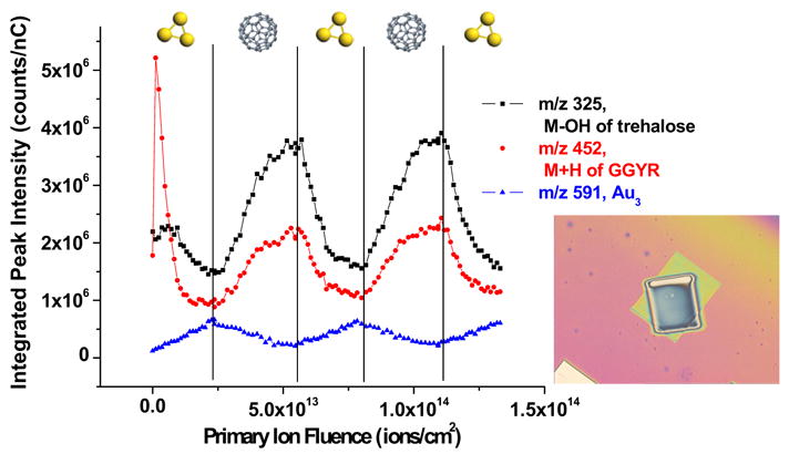
The sputtering properties of two representative cluster ion beams in secondary ion mass spectrometry (SIMS), C(60)(+) and Au(3)(+), have been directly compared. Organic thin films consisting of trehalose and dipalmitoylphosphatidylcholine (DPPC) are employed as prototypical targets. The strategy is to make direct comparison of the response of a molecular solid to each type of the bombarding cluster by overlapping the two ion beams onto the same area of the sample surface. The ion beams alternately erode the sample while keeping the same projectile for spectral acquisition. The results from these experiments are important to further optimize the use of cluster projectiles for SIMS molecular depth profiling experiments. For example, Au(3)(+) bombardment is found to induce more chemical damage as well as Au implantation when compared with C(60)(+). Moreover, C(60)(+) is found to be able to remove the damage and the implanted Au effectively. Discussions are also presented on strategies of enhancing sensitivity for imaging applications with cluster SIMS.


Figures

Figure 1
Figure 1
Secondary ion signal intensities are plotted against the primary ion dose during the alternative bombardment by Au3+ and C60+. The order of the projectile that is used to sputter for each turn is indicated on the figure. The inset is an optical image of the film after the overlap depth profile experiment. The smaller crater is created by C60+ sputtering while the bigger crater on the outside is created by Au3+ sputtering. All of the spectra are taken from the overlapped center area. The field of view is 1360 μm × 1020 μm.
Figure 2
Figure 2
(a) Plot of the ratio between m/z 437 to m/z 452 (M + H of GGYR) versus accumulated total ion dose. (b) XPS image of Au (Au 4f in red) and C (C1s in green) from part of the crater after extensive bombardment of Au3+. The field of view is 800 μm × 800 μm.
Figure 3
Figure 3
Plot of the ratio between m/z 19 to m/z 18 versus accumulated total ion dose.
Figure 4
Figure 4
Depth profile plot of the DPPC molecular ion M + H+ (m/z = 735) and principle DPPC headgroup fragments phosphocholine C5H15NPO4+(m/z=184) and choline C5H12N+ (m/z = 86)secondary ion signal intensities versus the primary ion dose accumulated during alternative bombardment with Au3+ and C60+. The sputter projectile used over a specified primary ion dose is illustrated in the figure.
Figure 5
Figure 5
Plots of the ratio between the fragmented choline ion C5H12N+ (m/z = 86) and the phosphocholine ion C5H15NPO4+(m/z=184) and the ratio between H3O+ (m/z = 19) and H2O+ (m/z = 18) (as a measure of surface protons accumulated during the depth profile) versus primary ion dose accumulated during alternative bombardment with Au3+ and C60+.
Figure 6
Figure 6
Depth profile plot of DPPC molecular fragments phosphocholine C5H15NPO4+(m/z=184) and choline C5H12N+ (m/z = 86) secondary ion intensities versus primary ion dose. Solid symbols represent secondary ion intensities acquired during a depth profile using C60+ only (as both the sputter source and the analysis source) and open symbols represent secondary ion intensities acquired during a depth profile using Au3+ only. The inset is an AFM image of a dry, porous DPPC film bombarded with C60+ at 200 × 200 μm2 field of view. Image is taken at a 400 × 400 μm2 field of view and indicates the film is 1000 nm thick with a 100 nm surface roughness.

References

    1. Jones EA, Lockyer NP, Vickerman JC. Mass Spectral Analysis and Imaging of Tissue by TOF-SIMS—the Role of Buckminsterfullerence, C60+, Primary Ions. Int J Mass Spectrom. 2006 in press.
    1. Winograd N. The Magic of Cluster SIMS. Anal Chem. 2005;77:142A–149A.
    1. Winograd N, Postawa Z, Cheng J, Szakal C, Kozole J, Garrison BJ. Improvements in SIMS Continue. Is the End in Sight? Appl Surf Sci. 2006;252(19):6836–6843.
    1. Cornett D, Lee T, Mahoney J. Matrix-Free Desorption of Biomolecules Using Massive Cluster-Impact. Rapid Commun Mass Spectrom. 1994;8:996–1000. - PubMed
    1. Tempez A, Schultz JA, Della-Negra S, Depauw J, Jacquet D, Novikov A, Lebeyec Y, Pautrat M, Caroff M, Ugarov M, Bensaoula H, Gonin M, Fuhrer K, Woods A. Orthogonal Time-of-Flight Secondary Ion Mass Spectrometric Analysis of Peptides Using Large Gold Clusters as Primary Ions. Rapid Commun Mass Spectrom. 2004;18:371–376. - PubMed

Publication types

MeSH terms