Review
. 2006 Dec 29;361(1476):2155-72.
doi: 10.1098/rstb.2006.1937.

Behavioural and neurophysiological evidence for face identity and face emotion processing in animals

Affiliations
Review

Behavioural and neurophysiological evidence for face identity and face emotion processing in animals

Andrew J Tate et al. Philos Trans R Soc Lond B Biol Sci. .

Abstract

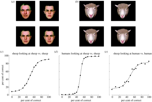
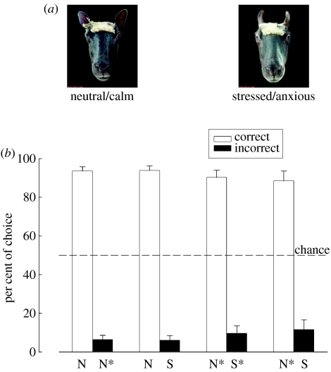
Visual cues from faces provide important social information relating to individual identity, sexual attraction and emotional state. Behavioural and neurophysiological studies on both monkeys and sheep have shown that specialized skills and neural systems for processing these complex cues to guide behaviour have evolved in a number of mammals and are not present exclusively in humans. Indeed, there are remarkable similarities in the ways that faces are processed by the brain in humans and other mammalian species. While human studies with brain imaging and gross neurophysiological recording approaches have revealed global aspects of the face-processing network, they cannot investigate how information is encoded by specific neural networks. Single neuron electrophysiological recording approaches in both monkeys and sheep have, however, provided some insights into the neural encoding principles involved and, particularly, the presence of a remarkable degree of high-level encoding even at the level of a specific face. Recent developments that allow simultaneous recordings to be made from many hundreds of individual neurons are also beginning to reveal evidence for global aspects of a population-based code. This review will summarize what we have learned so far from these animal-based studies about the way the mammalian brain processes the faces and the emotions they can communicate, as well as associated capacities such as how identity and emotion cues are dissociated and how face imagery might be generated. It will also try to highlight what questions and advances in knowledge still challenge us in order to provide a complete understanding of just how brain networks perform this complex and important social recognition task.


Figures

Figure 1
Figure 1
(a) Example of human face pictures that sheep (n=8) learned to discriminate between and below the same pictures morphed so that the difference between them is reduced to 10%; (b) same as (a) but for sheep face pictures and these were also tested on 10 human subjects to provide a comparison; (c) discrimination accuracy curve plotted for sheep discriminating between different degrees of morphing between the two images; (d) same but for humans looking at sheep and (e) for the same sheep looking at humans. In all the cases, 70% choice is considered significant (p<0.05).
Figure 2
Figure 2
(a) Pair of sheep faces presented during operant discrimination tasks. An example of (i) neutral/calm sheep face and the same individual displaying (ii) stress/anxiety is shown. During face identity recognition tasks, pairs of neutral/calm faces of two different conspecifics were presented. (b) Performance during face identity and face emotion recognition tasks. Sheep are able to recognize individual conspecifics by their faces ((i) N, neutral/calm face of a familiar conspecific; N*, neutral/calm face of an unfamiliar individual). Furthermore, sheep are able to discriminate between calm and stressed/anxious facial displays of familiar conspecific (N versus S). When presented with the same choice, however, using pictures of unfamiliar conspecifics (N* versus S*), sheep prefer the neutral display. When eventually presented with a choice between unfamiliar neutral faces and familiar stressed/anxious faces ((ii) N* versus S), sheep prefer the unfamiliar neutral faces.
Figure 3
Figure 3
(a) Picture set used to investigate response specificities of cells responding to a sheep face. (b) Histograms show overall mean±s.e.m. firing rate changes (per cent change from period with fixation spot displayed immediately before face stimulus is displayed) for 14 view-dependent (frontal view only) and 21 view-independent (equivalent responses to front and profile views) neurons recorded from the right temporal cotex of four sheep. (c) Same as (b) but for eight view-dependent and 11 view-independent neurons from the left temporal cortex.
Figure 4
Figure 4
(a) Histograms show proportions of view-dependent and view-independent face responsive neurons recorded in the temporal (n=69 cells from four sheep) and medial frontal (n=57 cells from five sheep) cortices. (b) Mean±s.e.m. latencies and rise times of 20 neurons in the right frontal cortex responding to the different views or faces in stimulus set shown in figure 3a.
Figure 5
Figure 5
Activity maps: pseudo-colour grids showing activity changes across the recording array during face discrimination tasks. (a) Example of different activation profiles in response to either a novel, non-familiar face or a learnt, familiar face. Overall, the number of units responding to the face decreases as the face becomes more familiar to the subject. Individually, some units no longer react to the stimulus while others show increases/decreases in their firing rates. (b) Activity map across grid in response to familiar face showing higher levels of inhibition as opposed to excitation across the population.
Figure 6
Figure 6
(a) Differential activity map of temporal cortical neurons during a face-based emotion recognition (FaBER) task. The map highlights the activity differences across the population observed in response to an emotional (stressed/anxious) face stimulus of a familiar sheep face as opposed to an unfamiliar sheep face. (b) In both hemispheres, the total number of neurons (T) responding to the face does not change irrespective of whether a familiar (N) or an unfamiliar (N*) neutral/calm sheep face is presented during the discrimination task. However, the proportion of E-type neurons (i.e. neurons with an increased firing rate, E) is higher whereas the number of I-type responses (decreased firing rate, I) is lower across the population if the neutral/calm face is unfamiliar (N*). (c) Bihemispherical comparison of the response latencies (ΔtL−R) of the neurons reveals right hemisphere dominance during sole face identity recognition tasks where animals discriminated between a familiar neutral/calm (N) and an unfamiliar neutral/calm (N*) face. Right hemisphere dominance is less pronounced during the face emotion recognition tasks (N versus S, N* versus S).
Figure 7
Figure 7
Mean±s.e.m. per cent change in firing rate in frontal cortex neurons in five maternal sheep responding to the sight of their lamb's face and to its odour. View-dependent neurons (n=12) only respond to a frontal view and not to the lamb's odour. However, view-independent neurons (n=25) respond equivalently to frontal and profile views and also to just the lamb's odour. For 8 of these cells when the lamb's face and odour were presented in combination there was no evidence for any additive response.

Similar articles

Cited by

References

    1. Aggleton J.P, Young A.W. The enigma of the amygdala: on its contribution to human emotion. In: Lane R.D, Nadel L, editors. Cognitive neuroscience of emotion. Oxford University Press; New York, NY: 2000. pp. 106–128.
    1. Baker C.I, Keysers C, Jellema T, Wicker B, Perrett D.I. Neuronal representation of disappearing and hidden objects in temporal cortex of the macaque. Exp. Brain. Res. 2001;140:375–381. doi:10.1007/s002210100828 - DOI - PubMed
    1. Bauer H.R, Philip M. Facial and vocal individual recognition in the common chimpanzee. Psychol. Rec. 1983;33:161–170.
    1. Baylis G.C, Rolls E.T, Leonard C.M. Selectivity between faces in the responses of a population of neurons in the cortex in the superior temporal sulcus of the monkey. Brain. Res. 1985;342:91–102. doi:10.1016/0006-8993(85)91356-3 - DOI - PubMed
    1. Bekoff M. Animal reflections. Nature. 2002;419:255. doi:10.1038/419255a - DOI - PubMed

Publication types

MeSH terms

LinkOut - more resources