. 2007 Mar 1;109(5):1897-907.
doi: 10.1182/blood-2006-08-044156. Epub 2006 Nov 21.

Retroviral vector insertion sites associated with dominant hematopoietic clones mark "stemness" pathways

Affiliations

Retroviral vector insertion sites associated with dominant hematopoietic clones mark "stemness" pathways

Olga S Kustikova et al. Blood. .

Abstract

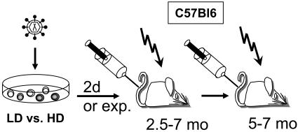
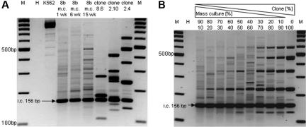
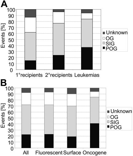
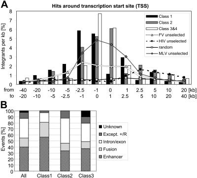
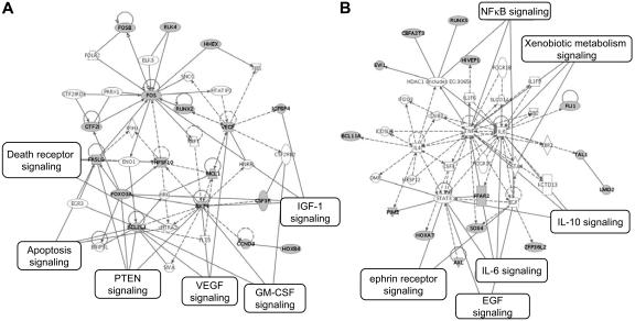
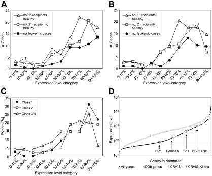
Evidence from model organisms and clinical trials reveals that the random insertion of retrovirus-based vectors in the genome of long-term repopulating hematopoietic cells may increase self-renewal or initiate malignant transformation. Clonal dominance of nonmalignant cells is a particularly interesting phenotype as it may be caused by the dysregulation of genes that affect self-renewal and competitive fitness. We have accumulated 280 retrovirus vector insertion sites (RVISs) from murine long-term studies resulting in benign or malignant clonal dominance. RVISs (22.5%) are located in or near (up to 100 kb [kilobase]) to known proto-oncogenes, 49.6% in signaling genes, and 27.9% in other or unknown genes. The resulting insertional dominance database (IDDb) shows substantial overlaps with the transcriptome of hematopoietic stem/progenitor cells and the retrovirus-tagged cancer gene database (RTCGD). RVISs preferentially marked genes with high expression in hematopoietic stem/progenitor cells, and Gene Ontology revealed an overrepresentation of genes associated with cell-cycle control, apoptosis signaling, and transcriptional regulation, including major "stemness" pathways. The IDDb forms a powerful resource for the identification of genes that stimulate or transform hematopoietic stem/progenitor cells and is an important reference for vector biosafety studies in human gene therapy.


Figures

Figure 1
Figure 1
Experimental setup of murine BMT studies using donor cells modified with different retroviral vectors. The enhancer-promoter contained in the long terminal repeat (LTR), the cDNA encoded by the vector, and the 3′ untranslated region (3′ UTR) are indicated in Table 2. LD indicates low dose of retroviral vector; HD, high dose; and exp, expansion in vivo.
Figure 2
Figure 2
LMPCR validation. (A) DNA of K562 mass cultures and cell clones containing different numbers of retroviral insertions was subjected to insertion site amplification by LMPCR using the conditions described in “Material and methods.” In contrast to the clonal DNA, mass culture DNA does not reveal dominant bands except when cells were propagated for several weeks, revealing a clonal imbalance. (B) Mixing mass culture DNA with increasing amounts of DNA from clone 2.4 reveals that LMPCR recovers dominant bands if these contribute greater than 70% of the population.
Figure 3
Figure 3
Retroviral vector insertion site (RVIS) distribution according to gene classes and type of transgene. (A) RVISs in known proto-oncogenes (POGs) increase in frequency over serial BMT and are most pronounced in leukemic clones. (B) No major impact of the transgene class was found except when the vector encoded a potent oncogene (TAg), which increased the probability to select for RVIS in POGs. SIGs indicates signaling genes; OGs, other genes.
Figure 4
Figure 4
Type of mutations. Data are shown with respect to gene class 1 (common insertion sites, proto-oncogenes, and self-renewal genes), class 2 (signaling genes), and classes 3 and 4 (other and unknown genes). (A) Position of RVIS in the Insertional Dominance Database (IDDb) around the transcriptional start site. Reference data insertion sites of different vectors in freshly transduced cells, shown as lines, were kindly provided by G. Trobridge and D. Russell. MLV indicates murine leukemia virus vector; FV, foamy virus vector; HIV, human immunodeficiency virus vector; random, computer-predicted random insertion pattern. (B) Overrepresentation of enhancer mutations in class 1 genes. RVISs were analyzed for the different types of retroviral insertional mutations proposed earlier. Insertions located downstream but in an antisense orientation do not correspond to the definition of enhancer mutations suggested in Uren et al and are therefore labeled “Except. +/R.”.
Figure 5
Figure 5
Ingenuity analyses of the genes listed in Table 3 reveal 2 major pathways. Note that further members of these pathways (A-B) may be highlighted when extending the analysis to the full IDDb. That is, Siva shown on the bottom of Figure 5A is a chromosomal neighbor of Akt1; this locus represents a CRVIS in the IDDb (Table 3; Table S1).
Figure 6
Figure 6
The probability of retroviral vector insertion but not the probability of forming a common insertion site depends on the expression level of the affected gene. (A) Array data from enriched hematopoietic progenitors containing both long-term and short-term repopulating cells (LSK cells, freshly isolated) were divided into 10 equal bins according to relative gene expression levels. The curves show the number of genes marked by RVISs in the different bins. Irrespective of the selection conditions (primary recipient, secondary recipient, or leukemia), the probability of RVIS is highest in the 40% most highly expressed genes. (B) Similar results were obtained when examining array data from LSK cells that were cultured for 2 days. (C) The selection for insertions in highly expressed genes is most pronounced for class 1 genes. (D) Expression levels of all genes detected by the arrays of LSK cells versus all RVIS genes of the IDDb, showing that the latter clearly have a much higher expression. The CRVIS genes of the IDDb are superimposed, showing that these do not cluster in the highest expression levels. Labeled genes represent CRVISs that were hit 3 times or more. Genes below the dotted line are not expressed in LSK cells.

References

    1. Mikkers H, Berns A. Retroviral insertional mutagenesis: tagging cancer pathways. Adv Cancer Res. 2003;88:53–99. - PubMed
    1. Akagi K, Suzuki T, Stephens RM, Jenkins NA, Copeland NG. RTCGD: retroviral tagged cancer gene database. Nucleic Acids Res. 2004;32:D523–D527. - PMC - PubMed
    1. Li Z, Dullmann J, Schiedlmeier B, et al. Murine leukemia induced by retroviral gene marking. Science. 2002;296:497. - PubMed
    1. Kustikova OS, Fehse B, Modlich U, et al. Clonal dominance of hematopoietic stem cells triggered by retroviral gene marking. Science. 2005;308:1171–1174. - PubMed
    1. Modlich U, Kustikova O, Schmidt M, et al. Leukemias following retroviral transfer of multidrug resistance 1 are driven by combinatorial insertional mutagenesis. Blood. 2005;105:4235–4246. - PubMed

Publication types