Review
. 2006 Dec;28(12):1181-93.
doi: 10.1002/bies.20502.

The molecular basis of neurosensory cell formation in ear development: a blueprint for hair cell and sensory neuron regeneration?

Affiliations
Review

The molecular basis of neurosensory cell formation in ear development: a blueprint for hair cell and sensory neuron regeneration?

Bernd Fritzsch et al. Bioessays. 2006 Dec.

Abstract

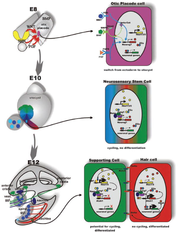
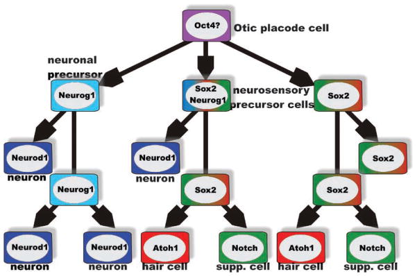
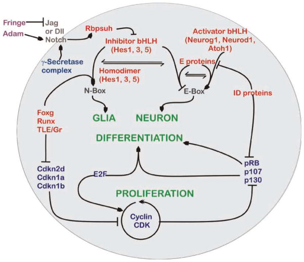
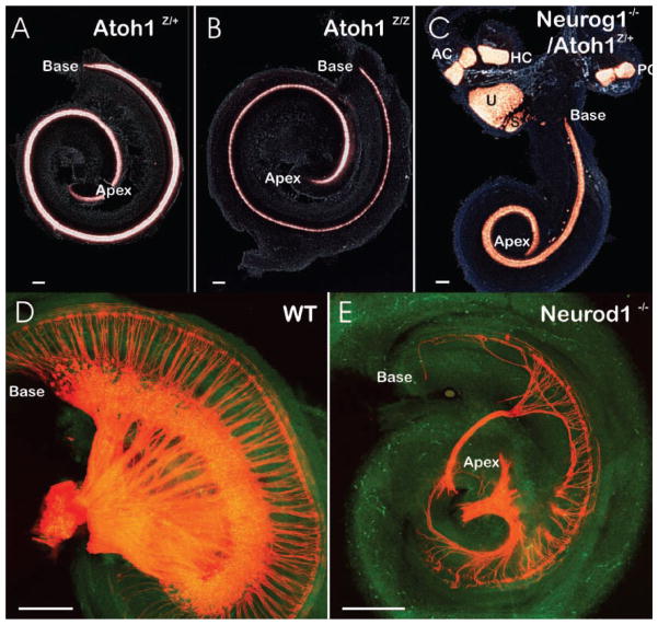
The inner ear of mammals uses neurosensory cells derived from the embryonic ear for mechanoelectric transduction of vestibular and auditory stimuli (the hair cells) and conducts this information to the brain via sensory neurons. As with most other neurons of mammals, lost hair cells and sensory neurons are not spontaneously replaced and result instead in age-dependent progressive hearing loss. We review the molecular basis of neurosensory development in the mouse ear to provide a blueprint for possible enhancement of therapeutically useful transformation of stem cells into lost neurosensory cells. We identify several readily available adult sources of stem cells that express, like the ectoderm-derived ear, genes known to be essential for ear development. Use of these stem cells combined with molecular insights into neurosensory cell specification and proliferation regulation of the ear, might allow for neurosensory regeneration of mammalian ears in the near future.


Figures

Figure 1
Figure 1
Organ, cell and molecular interactions in ear development. The morphogenesis (left) and some molecular interactions underlying proliferation and cell fate decision (right) are depicted in this scheme. Morphogenesis transforms a small patch of ectoderm between embryonic days 8 and 12 into a complex labyrinth of ducts and recesses that harbors the six sensory epithelia of the mammalian ear in strategic positions for extraction of epithelia-specific energy. Delamination of sensory neurons generates the vestibular and cochlear sensory neurons that connect specific sensory epithelia of the ear to specific targets in the hindbrain. One of the earliest steps in this process is the selection of otic placode cells through the interaction of several diffusible factors; in particular, FGF and WNT signaling upregulates both inhibitory and activating bHLH genes to switch the cell fate through downregulation of BMP signaling, specifying the position and size of the otic placode (top right). These stem cells will, through the interaction of activator- and inhibitor-type bHLH genes remain in cycling phase without differentiation resulting in clonal expansion. As cells progress through the cycles, they will change their fate determination, giving rise to neurosensory stem cells (middle right) that form by asymmetric divisions all sensory neurons of the ear. Some neurosensory stem cells as well as independently arising cells of the otic placode turn into sensory epithelia precursor cells (SNP). These cells will give rise by asymmetric divisions to hair cells and supporting cells (bottom right). Exit from the cell cycle, combined with proper cell fate specification to, eg hair cell and supporting cell, will be mediated in part by the NOTCH-reinforced switch to either explosive upregulation of proneuronal bHLH genes (Atoh1 in the case of hair cells) or of inhibitory bHLH genes (such as Hes1 or Hes5) by the γ-secretase-cleaved Notch fragment that binds to RBPSUH (formerly Rbp-J). The action of HES homodimers on N-boxes to turn on proneuronal genes is enhanced through interaction with the TLE, RUNX, FOXG and genes. Consequently, eliminating for example Foxg1 results in diminished efficacy of HES signaling resulting in premature cell cycle exit and differentiation. Shortly after E14, all proliferative activity in the PNP progenitors stops and no new sensory neurons or hair cells will form. Modified after Refs ,.
Figure 2
Figure 2
Cell-type-specific and overlapping precursors. Analysis of several null mutations suggest that there is an initial formation of two, partially overlapping, precursor populations, a neuronal precursor characterized by Neurog1 expression and a neurosensory precursor, characterized by Sox2 expression. The 40–80% reduction of hair cell and supporting cell formation in Neurog1 null mice suggests that the size of the common neuronal/neurosensory precursor population varies in different sensory epithelia. The later-expressed bHLH gene Neurod1 does not show this massive effect on hair cells and appears to be exclusively expressed in differentiating neurons. Absence of hair cell differentiation in Sox2 and Atoh1 null mice suggests that these genes are essential for hair cell formation, no matter what origin. Supporting cells depend on the hair-cell-mediated upregulation of Notch (and Hes) for their differentiation and will turn into hair cells in the absence of proper Notch/Hes signaling. Modified after Refs ,,,,.
Figure 3
Figure 3
Signaling pathways for inner ear proliferation and differentiation. This schematic diagram represents an overview of the known and presumed interactive pathways for proliferation and differentiation of the neurosensory cells in the inner ear. Signaling of the membrane-bound (brown) Notch receptors by binding to their ligands, Delta and Jagged, can be influenced by the extracellular (purple) Fringe and ADAM enzymes. Fringe inhibits (blocked line) the Notch binding of Jagged, while Adam cleaves the Notch receptor to potentate its activation (lined arrow). The cleavage of intercellular domain fragment of Notch is done by the cytoplasmic (dark blue) γ-secretase complex which then activates the nuclear protein (red) RBPSUH. Inactivated RBPSUH blocks transcription of the Hes genes whereas activation enhances transcription. Homodimers of HES proteins can bind to N-boxes to initiate differentiation (green) of glial precursors. N-box binding of HES homodimers is regulated further by a FOXG, RUNX and TLE promoter complex. Heterodimers between HES and E proteins bind and competitively block usage of E-box-binding sites. Activation of E-box promoter sequences is through the combined E-protein and the activator bHLH heterodimers and this permits neuronal differentiation. To do this, the activator bHLH proteins compete with HES proteins for the E-protein-binding partners. E proteins can also be inactivated from DNA binding through interaction with the inhibitor of DNA-binding (ID) proteins, which also suppress the cell cycle (blue) retinoblastoma isoforms. The pRB isoforms alone or in combination E2F proteins cause cell differentiation. Cell proliferation (green) is mediated through the proteins of cyclin CDK pathway that phosphorylate Rb to allow E2F proteins to initiate the S-phase entry. The cyclin CDK proteins can also inhibit differentiation via pRB phosphorylation, whereas cyclin-dependent kinase inhibitors (Cdkn) prevent proliferation. Expression of the CDKNs is blocked by the FOXG, RUNX and TLE complex, allowing differentiation of glia cells through enhanced action of HES homodimers on the N-Box. Modified after Refs ,,,.
Figure 4
Figure 4
Examples of gene effects on histogenesis and morphogenesis. A,B,D,E: Flat-mounted cochlea or C: entire ears show the effects of targeted deletion of an activator-type bHLH gene (Atoh1, B; Neurog1, C; Neurod1, E) on the presence of hair cells (revealed by Atoh1–lac Z expression in A–C) or innervation (revealed by lipophilic dye tracing in D,E). Note that both the distribution of Atoh1–lac Z-positive cells as well as the overall length of the cochlea (base and apex are indicated) show little difference in Atoh1–lac Z heterozygote and null mutants, despite the fact that no hair cells differentiate in Atoh1 null mice. This suggests that the late upregulation of a bHLH gene in cells destined to exit the cell cycle is of little consequence for morphogenesis and cellular patterning in the ear. In contrast, earlier upregulated bHLH genes such as Neurog1 (C) or Neurod1 (E) have a more profound morphogenetic effect such as shortening of the cochlea (C,E) or almost complete loss of sensory epithelia (saccule in E). Additional effects are displaced development of some hair cells outside the typical sensory epithelia (C) or loss of a large fraction of sensory neurons combined with an alteration in the pattern of innervation. Modified after Refs ,, AC, anterior crista; HC, horizontal crista; PC, posterior crista, S, saccule; U, utricle. Bar indicates 100 μm.
Figure 5
Figure 5
bHLH gene interactions in retinal ganglion cell specification. The most-detailed single-cell quantitative PCR analysis shows that relative concentrations of bHLH transcripts vary systematically during chicken retina ganglion cell formation. In the first phase (red line), Hes1 transcript exceeds that of Neurog2 and very much that of Atoh7. This dominance of inhibitory bHLH gene expression will result in homodimers on N-boxes (yellow hexagons) as well as few heterodimers of Neurog2 with E2a on E-boxes (lilac/blue hexagons). In phase 2 (blue lines) Hes1 is downregulated allowing Atoh7 transcript to become as prominent as Neurog2 and to form heterodimers with E2a proteins to bind to specific E-boxes (red and blue hexagons). In the third phase (green line) Atoh7 is further upregulated to drive ganglion cell differentiation as well as preventing the developing neuron from reentering the cell cycle. Modified after Refs .

References

    1. Rakic P. Neuroscience. No more cortical neurons for you. Science. 2006;313:928–929. - PubMed
    1. Rask-Andersen H, Bostrom M, Gerdin B, Kinnefors A, Nyberg G, et al. Regeneration of human auditory nerve. In vitro/in video demonstration of neural progenitor cells in adult human and guinea pig spiral ganglion. Hear Res. 2005;203:180–191. - PubMed
    1. Li H, Liu H, Heller S. Pluripotent stem cells from the adult mouse inner ear. Nat Med. 2003;9:1293–1299. - PubMed
    1. Fritzsch B, Tessarollo L, Coppola E, Reichardt LF. Neurotrophins in the ear: their roles in sensory neuron survival and fiber guidance. Prog Brain Res. 2004;146:265–278. - PubMed
    1. Sugawara M, Corfas G, Liberman MC. Influence of supporting cells on neuronal degeneration after hair cell loss. J Assoc Res Otolaryngol. 2005;6:136–147. - PMC - PubMed

Publication types