. 2006 Nov;3(11):e441.
doi: 10.1371/journal.pmed.0030441.

Complement lysis activity in autologous plasma is associated with lower viral loads during the acute phase of HIV-1 infection

Affiliations

Complement lysis activity in autologous plasma is associated with lower viral loads during the acute phase of HIV-1 infection

Michael Huber et al. PLoS Med. 2006 Nov.

Abstract

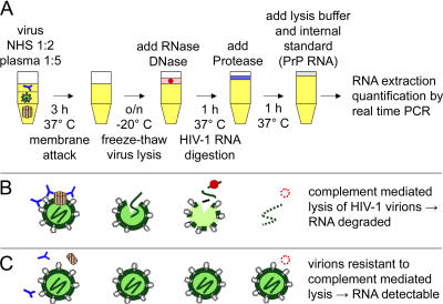
Background: To explore the possibility that antibody-mediated complement lysis contributes to viremia control in HIV-1 infection, we measured the activity of patient plasma in mediating complement lysis of autologous primary virus.

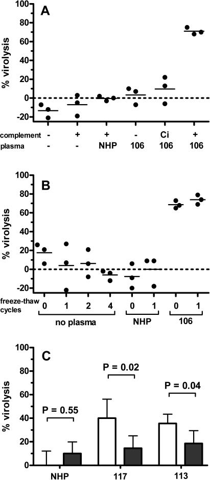
Methods and findings: Sera from two groups of patients-25 with acute HIV-1 infection and 31 with chronic infection-were used in this study. We developed a novel real-time PCR-based assay strategy that allows reliable and sensitive quantification of virus lysis by complement. Plasma derived at the time of virus isolation induced complement lysis of the autologous virus isolate in the majority of patients. Overall lysis activity against the autologous virus and the heterologous primary virus strain JR-FL was higher at chronic disease stages than during the acute phase. Most strikingly, we found that plasma virus load levels during the acute but not the chronic infection phase correlated inversely with the autologous complement lysis activity. Antibody reactivity to the envelope (Env) proteins gp120 and gp41 were positively correlated with the lysis activity against JR-FL, indicating that anti-Env responses mediated complement lysis. Neutralization and complement lysis activity against autologous viruses were not associated, suggesting that complement lysis is predominantly caused by non-neutralizing antibodies.

Conclusions: Collectively our data provide evidence that antibody-mediated complement virion lysis develops rapidly and is effective early in the course of infection; thus it should be considered a parameter that, in concert with other immune functions, steers viremia control in vivo.


Conflict of interest statement

Competing Interests: The authors declare that no competing interests exist.

Figures

Figure 1
Figure 1. Schematic Overview of the Virion Lysis Assay
(A) Primary HIV-1 virions were incubated with autologous plasma and complement and freeze-thawed once, and viral RNA was digested by RNase and DNase. After inactivation of RNase and DNase by protease and addition of an internal standard (PrP RNA), RNA was extracted and quantified by real-time PCR. (B) HIV-1 virions sensitive to antibody-mediated complement lysis were disrupted, making viral RNA accessible for degradation. (C) Viral RNA of complement lysis-resistant virions remained intact. RNA was extracted and could be quantified by real-time PCR.
Figure 2
Figure 2. Virolysis by Antibody and Complement
(A) Plasma from HIV-positive individuals specifically lyses HIV in presence of active complement. Virus isolate JR-FL was incubated with complement (+), without complement (−), or with inactivated complement (Ci) either in the absence of plasma (−) or with plasma from uninfected individuals (NHP) or from patient 106. One of three independent experiments is shown. (B) Up to four freeze-thaw cycles do not destroy intact HIV-1 virions. Virus JR-FL was incubated in the presence of active complement with medium (no plasma), with plasma from uninfected persons (NHP), or with plasma from patient 106. Reaction mixtures were subjected to the indicated number of freeze-thaw cycles and the effect on virus disintegration was measured. One of two independent experiments is shown. (C) IgG depletion reduces lysis activity. Plasma of uninfected individuals (NHP) and of patients 117 and 113 were depleted of IgG with Protein G Sepharose beads (darkened bars) and lysis activity was compared to untreated plasma (open bars). Error bars indicate standard deviation of triplicate measurements. Groups were compared using Mann-Whitney U test.
Figure 3
Figure 3. Lysis Activity against Virus in Plasma of Acutely and Chronically Infected Patients
Complement-mediated lysis activity against autologous (A) and heterologous (JR-FL) (B) virus was compared between acutely (blue triangles) and chronically (green circles) infected individuals. HIV-specific lysis activity is present at acute and chronic diseases stages but generally higher during chronic infection. Crosses denote uninfected plasma controls. Groups were compared using Mann-Whitney U test.
Figure 4
Figure 4. Correlation Analysis of Antibody Responses and Virolysis Activity
(A) Correlation analysis of anti-gp120 and anti-gp41 antibody titers in plasma samples from the acute and chronic patient groups shows that titers to the envelope glycoprotein correlate tightly independently of disease stage. (B–D) Correlation analyses of heterologous lysis (JR-FL) with anti-gp120 (B) or anti-gp41 (C) revealed positive correlations, whereas anti-p24 (D) antibody titers did not associate with heterologous lysis. This suggests that lysis activity is driven by envelope-specific antibodies. Blue triangles denote acutely, green circles denote chronically infected patients. Spearman's rank correlation coefficient (rho) and p-values are depicted for the entire cohort (all), the acute patients (a), and the chronic patients (c). If antibody titers were below 1, the value 1 was used for statistical analysis.
Figure 5
Figure 5. Longitudinal Assessment of Autologous Complement Lysis Activity in Patient Plasma
Lysis activity against autologous virus (red circles) of plasma from six acutely HIV-1-infected patients was measured longitudinally and plotted against anti-gp120 titers (orange triangles), anti-gp41 titers (blue squares), and viral load (asterisks). At the first data point, patients were treatment-naïve and acutely HIV infected. All patients (except patient 022) subsequently went on antiretroviral therapy for the indicated time periods. Time point 0 was assigned to the date of treatment interruption and the remaining time points were calculated according to this time point. (A) Patient 022 (treatment-naïve), (B) patient 015, (C) patient 003, (D) patient 016, (E) patient 026, and (F) patient 002.
Figure 6
Figure 6. Neutralizing Antibodies Are Not the Major Constituent of Complement Lysis-Inducing Antibodies
Complement lysis activity of non-neutralizing and neutralizing patient plasmas was compared in the acute and the chronic infection cohorts. Autologous neutralization (A and B) and heterologous (JR-FL) (C and D) neutralization are shown, with blue triangles denoting acutely, green circles chronically infected patients. Each data point represents the mean of two or three independent experiments with plasma from the same patient. Groups were compared using Mann-Whitney U test.
Figure 7
Figure 7. Influence of Complement on Viral Infectivity In Vitro
Inhibitory activity of patient plasma against the heterologous virus JR-FL was measured in presence of active (C+) or heat-inactivated (Ci) complement on TZM-bl cells. Blue triangles denote acutely, green circles chronically infected patients. Data points are means of two independent experiments with plasma from the same patient. Differences in inhibition between inactivated and active complement within the subgroups were compared using Wilcoxon signed-rank test. The results demonstrate that complement increases the inhibitory activity of HIV specific antibodies in vitro.
Figure 8
Figure 8. Correlation Analysis of Complement Virolysis and In Vivo Viremia Levels
In vitro-determined autologous virolysis activity was correlated with in vivo HIV-1 RNA copies measured per milliliter of plasma in the acutely infected group (A) and the chronically infected group (B). Correlations were evaluated using Spearman's rank correlation. Our data demonstrate that increased antibody-mediated complement lysis coincides with lower viral loads in the acute phase. No evidence was found for a similar correlation in the chronically infected group.

References

    1. Parren PW, Moore JP, Burton DR, Sattentau QJ. The neutralizing antibody response to HIV-1: Viral evasion and escape from humoral immunity. AIDS. 1999;13:S137–S162. - PubMed
    1. Mascola JR, Stiegler G, VanCott TC, Katinger H, Carpenter CB, et al. Protection of macaques against vaginal transmission of a pathogenic HIV-1/SIV chimeric virus by passive infusion of neutralizing antibodies. Nat Med. 2000;6:207. - PubMed
    1. Montefiori DC, Hill TS, Vo HTT, Walker BD, Rosenberg ES. Neutralizing antibodies associated with viremia control in a subset of individuals after treatment of acute human immunodeficiency virus type 1 infection. J Virol. 2001;75:10200–10207. - PMC - PubMed
    1. Wei X, Decker JM, Wang S, Hui H, Kappes JC, et al. Antibody neutralization and escape by HIV-1. Nature. 2003;422:307–312. - PubMed
    1. Richman DD, Wrin T, Little SJ, Petropoulos CJ. Rapid evolution of the neutralizing antibody response to HIV type 1 infection. Proc Natl Acad Sci U S A. 2003;100:4144–4149. - PMC - PubMed

Publication types