. 2006 Oct;2(10):e110.
doi: 10.1371/journal.ppat.0020110.

SabA is the H. pylori hemagglutinin and is polymorphic in binding to sialylated glycans

Affiliations

SabA is the H. pylori hemagglutinin and is polymorphic in binding to sialylated glycans

Marina Aspholm et al. PLoS Pathog. 2006 Oct.

Abstract

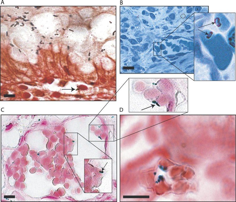
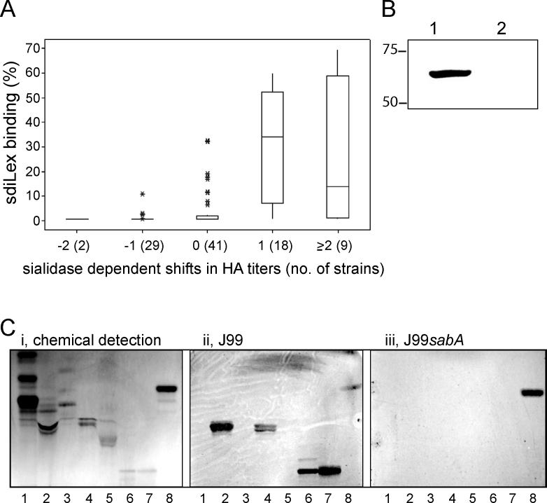
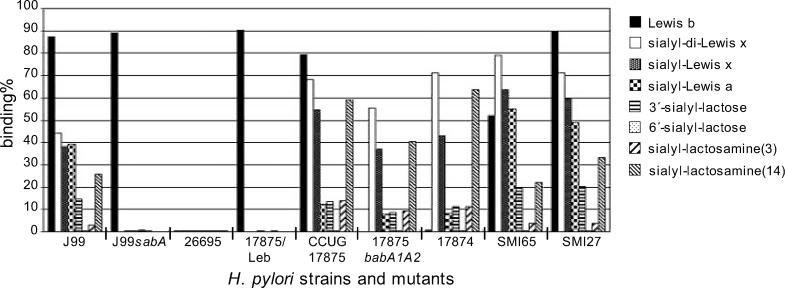
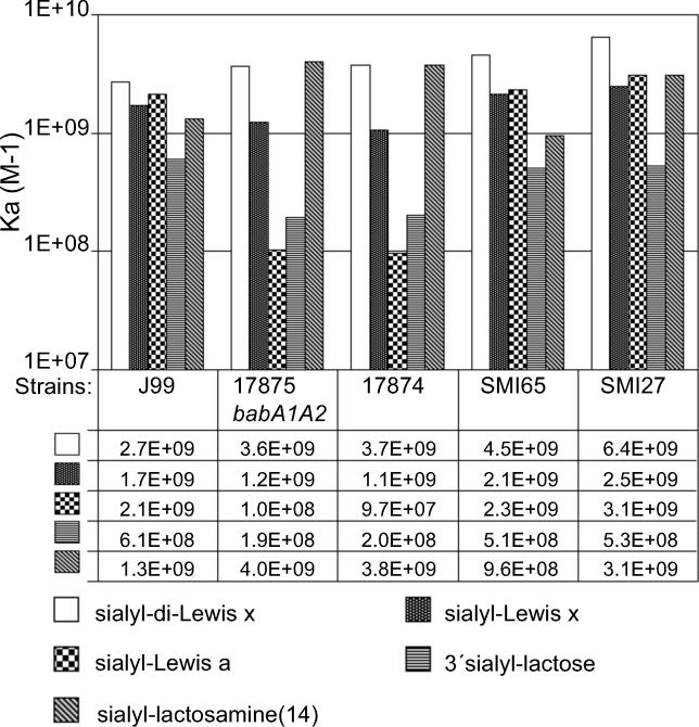
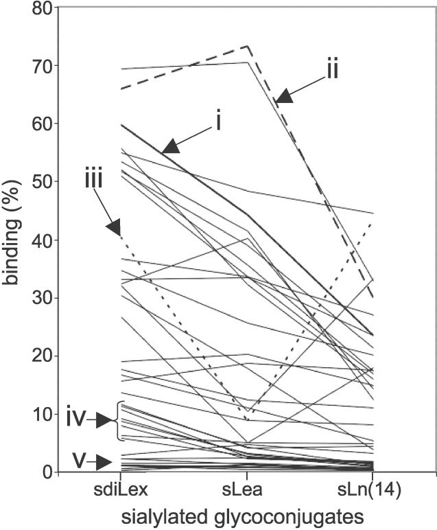
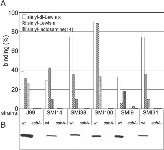
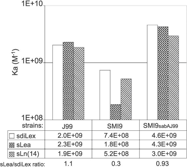
Adherence of Helicobacter pylori to inflamed gastric mucosa is dependent on the sialic acid-binding adhesin (SabA) and cognate sialylated/fucosylated glycans on the host cell surface. By in situ hybridization, H. pylori bacteria were observed in close association with erythrocytes in capillaries and post-capillary venules of the lamina propria of gastric mucosa in both infected humans and Rhesus monkeys. In vivo adherence of H. pylori to erythrocytes may require molecular mechanisms similar to the sialic acid-dependent in vitro agglutination of erythrocytes (i.e., sialic acid-dependent hemagglutination). In this context, the SabA adhesin was identified as the sialic acid-dependent hemagglutinin based on sialidase-sensitive hemagglutination, binding assays with sialylated glycoconjugates, and analysis of a series of isogenic sabA deletion mutants. The topographic presentation of binding sites for SabA on the erythrocyte membrane was mapped to gangliosides with extended core chains. However, receptor mapping revealed that the NeuAcalpha2-3Gal-disaccharide constitutes the minimal sialylated binding epitope required for SabA binding. Furthermore, clinical isolates demonstrated polymorphism in sialyl binding and complementation analysis of sabA mutants demonstrated that polymorphism in sialyl binding is an inherent property of the SabA protein itself. Gastric inflammation is associated with periodic changes in the composition of mucosal sialylation patterns. We suggest that dynamic adaptation in sialyl-binding properties during persistent infection specializes H. pylori both for individual variation in mucosal glycosylation and tropism for local areas of inflamed and/or dysplastic tissue.


Conflict of interest statement

Competing interests. In 2002 TB filed a patent application for the use of SabA as a vaccine candidate, International PCT pending number PCT/SE02/00301/ Helicobacter pylori sialic acid binding adhesin, SabA, and sabA gene.

Figures

Figure 1
Figure 1. H. pylori Adheres to Erythrocytes in Capillaries and Post-Capillary Venules of Infected Humans and Rhesus Monkeys
(A) Genta-stained section of human gastric biopsy. Black spiral- and comma-shaped bacteria are observed in the lumen of the stomach, adherent to gastric epithelial cells, within the mucus globule of the cells. Bacterial cells (arrow) are also present in close contact to an erythrocyte within a capillary located in the supporting connective tissue of lamina propria of the mucosa. (B) Section of human gastric biopsy stained with toluidine blue. A capillary vessel lined by endothelial cells is visible in the lamina propria of the mucosa. It contains several erythrocytes to which H. pylori are attached. Insert: higher magnification of two H. pylori (arrows) in close approximation to erythrocytes. (C) Section of a Rhesus monkey gastric biopsy. In situ hybridization was performed using probes specific for H. pylori 16S rRNA, demonstrating the presence of several H. pylori apparently attached to erythrocyte surfaces of a post-capillary venule located in the lamina propria of submucosa. Inserts: higher magnification of H. pylori bacterial cells (arrows) in close approximation to erythrocytes. (D) Section of a human gastric biopsy. In situ hybridization was performed using probes specific for H. pylori 16S rRNA. This high magnification of a capillary immediately adjacent to a gastric gland (on the top-right corner of the picture) demonstrates the presence of several H. pylori bacterial cells, stained blue, apparently attached to the surfaces of erythrocytes. Bars = 5 μm.
Figure 2
Figure 2. Characterization of Binding Properties of the SabA Adhesin, and Its siaHA Properties
(A) A panel of 99 Swedish clinical H. pylori isolates was tested for sia-HA properties and for binding to 125I-sdiLex conjugate. The numbers on the x axis indicate the shifts in HA titers after sialidase treatment: positive values indicate lowered sia-HA titers (i.e., sia-HA, whereas negative values indicate increased HA titers (i.e., sialic acid–independent HA). No change in HA titer is indicated by 0. The y axis gives the percentage of bound sLex-conjugate. (B) SabA was affinity-adsorbed to erythrocytes from a cell-surface protein extract of strain J99. Immunostaining using SabA antibodies confirmed the presence of SabA adsorbed onto the erythrocyte surfaces as a result of binding to sialylated glycans (lane 1), whereas SabA was completely absent when erythrocytes had been depleted of sialic acid by sialidase treatment prior to the test (lane 2). Molecular weight markers (in kDa) are indicated. (C) Binding of H. pylori strains J99 and J99sabA to human erythrocyte glycosphingolipids. (i) Chemical detection by anisaldehyde. (ii–iii) Autoradiograms obtained by binding of 35S-labeled H. pylori strain J99 and the J99sabA mutant, respectively, to separated glycosphingolipids. The lanes contain non-acid glycosphingolipids of human erythrocytes, 40 μg (lane 1); gangliosides of human erythrocytes, 40 μg (lane 2); GM3 ganglioside (NeuAcα2–3Galβ4Glcβ1Cer), 4 μg (lane 3); NeuAcα2–3-neolactotetraocylceramide (NeuAcα2–3Galβ4GlcNAcβ3Galβ4Glcβ1Cer), 4 μg (lane 4); NeuAcα2–6-neolactotetraocylceramide (NeuAcα2–6Galβ4GlcNAcβ3Galβ4Glcβ1Cer), 4 μg (lane 5); G-10 ganglioside (NeuAcα2–3Galβ4GlcNAcβ6 (NeuAcα2–3Galβ4GlcNAcβ3)Galβ4GlcNAcβ3Galβ4Glcβ1Cer), 1 μg (lane 6); G9-B ganglioside (Galα3(Fucα2)Galβ4GlcNAcβ6 (NeuAcα2–3Galβ4GlcNAcβ3)Galβ4GlcNAcβ3Galβ4Glcβ1Cer), 1 μg (lane 7); and reference gangliotriaosylceramide (GalNAcβ4Galβ4Glcβ1Cer) of mouse feces, 4 μg (lane 8).
Figure 3
Figure 3. Binding of H. pylori Strains and Clinical Isolates to Fucosylated and Sialylated Glycans of Various Complexities
H. pylori reference strains, mutants, and clinical isolates were tested for binding to the fucosylated Leb antigen and to a series of sialylated antigens, all presented by 125I-labeled albumin conjugates. The y axis gives the percentage of bound conjugate.
Figure 4
Figure 4. Binding Affinities Analyzed According to Scatchard of H. pylori Reference Strains, a babA Deletion Mutant, and Clinical Isolates for Sialylated Glycans
Figure 5
Figure 5. Different Sialyl-Dependent Binding Modes of H. pylori Identified by Use of sdiLex, sLea, and sLn Conjugates
A total of 39 Swedish clinical isolates were investigated for detailed sialyl-dependent binding properties. Representatives of the different binding modes for 125I-labeled sialylated glycans are illustrated in the diagram: (i) (in thick line) binds efficiently to all three sialylated glycans with preferential binding to sdiLex; (ii) binds to all three sialylated glycans, with better binding to sLea (“Λ-shaped” hatched line); (iii) binds preferentially to sdiLex and sLn(14) (“V-shaped” dotted line); (iv) binds preferentially to sdiLex but exhibits only modest binding for sLea and sLn(14) sialyl conjugates; and (v) binds modestly for all sialyl conjugates (<5% bound conjugate). The y axis gives the percentage of bound conjugate.
Figure 6
Figure 6. Analyses of SabA-Dependent Binding among Clinical Isolates
(A) Binding characteristics of H. pylori clinical isolates and SabA-deficient isogenic mutants were studied by use of soluble 125I-labeled sdiLex, sLea, and sLn(14) conjugates. (B) Immunoblot analysis of H. pylori clinical isolates and mutant strains using SabA antibodies. There were no SabA bands detected among the sabA deletion mutants; actually, there were no additional bands on the blots.
Figure 7
Figure 7. Analyses of Binding Affinities for Sialylated Antigens by SabA from Strain J99 Complementarily Expressed in Background Clinical Strain SMI9
Binding affinities of H. pylori strains J99, SMI9, and the complementation mutant SMI9sabAJ99 were analyzed according to Scatchard. Binding affinities and the percentage ratio of sLex/sLea binding affinities are indicated.

References

    1. Cover TL, Berg DE, Blaser MJ, Mobley HLT. Helicobacter pylori pathogenesis. In: Groisman EA, editor. Principles of bacterial pathogenesis. San Diego (California): Academic Press; 2001. pp. 509–558.
    1. Alm RA, Ling LS, Moir DT, King BL, Brown ED, et al. Genomic-sequence comparison of two unrelated isolates of the human gastric pathogen Helicobacter pylori. Nature. 1999;397:176–180. - PubMed
    1. Schreiber S, Konradt M, Groll C, Scheid P, Hanauer G, et al. The spatial orientation of Helicobacter pylori in the gastric mucus. Proc Natl Acad Sci U S A. 2004;101:5024–5029. - PMC - PubMed
    1. Emody L, Carlsson A, Ljungh A, Wadström T. Mannose-resistant haemagglutination by Campylobacter pylori. Scand J Infect Dis. 1988;20:353–354. - PubMed
    1. Unemo M, Aspholm-Hurtig M, Ilver D, Bergström J, Borén T, et al. The sialic acid binding SabA adhesin of Helicobacter pylori is essential for nonopsonic activation of human neutrophils. J Biol Chem. 2005;280:15390–15397. - PubMed

Publication types

MeSH terms