. 2007 Feb;81(3):1379-89.
doi: 10.1128/JVI.01712-06. Epub 2006 Nov 22.

E6AP-dependent degradation of DLG4/PSD95 by high-risk human papillomavirus type 18 E6 protein

Affiliations

E6AP-dependent degradation of DLG4/PSD95 by high-risk human papillomavirus type 18 E6 protein

Keisuke Handa et al. J Virol. 2007 Feb.

Abstract

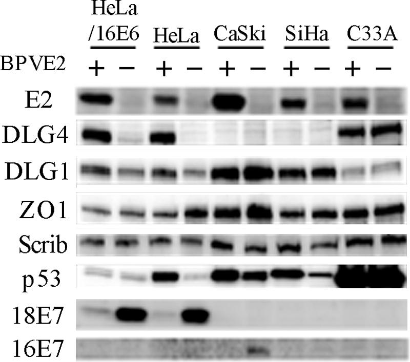
In most cervical cancers, DNAs of high-risk mucosotropic human papillomaviruses (HPVs), such as types 16 and 18, are maintained so as to express two viral proteins, E6 and E7, suggesting that they play important roles in carcinogenesis. The carboxy-terminal PDZ domain-binding motif of the E6 proteins is in fact essential for transformation of rodent cells and induction of hyperplasia in E6-transgenic mouse skin. To date, seven PDZ domain-containing proteins, including DLG1/hDLG, which is a human homologue of the Drosophila discs large tumor suppressor (Dlg), have been identified as targets of high-risk HPV E6 proteins. Here, we describe DLG4/PSD95, another human homologue of Dlg, as a novel E6 target. DLG4 was found to be expressed in normal human cells, including cervical keratinocytes, but only to a limited extent in both HPV-positive and HPV-negative cervical cancer cell lines. Expression of HPV18 E6 in HCK1T decreased DLG4 levels more strongly than did HPV16 E6, the carboxy-terminal motif of the proteins being critical for binding and degradation of DLG4 in vitro. DLG4 levels were restored by expression of either E6AP-specific short hairpin RNA or bovine papillomavirus type 1 E2 in HeLa but not CaSki or SiHa cells, reflecting downregulation of DLG4 mRNA as opposed to protein by an HPV-independent mechanism in HPV16-positive cancer lines. The tumorigenicity of CaSki cells was strongly inhibited by forced expression of DLG4, while growth in culture was not inhibited at all. These results suggest that DLG4 may function as a tumor suppressor in the development of HPV-associated cancers.


Figures

FIG. 1.
FIG. 1.
Expression of DLG4 in cervical cancer cell lines and primary human cells and the effects of various E6 proteins in HCK1T cells. (A) Expression levels of DLG4, DLG1, ZO1, Scrib, and p53 analyzed by immunoblotting. All cell proteins were extracted from subconfluent cultures. The same amounts of each cell extract (20 μg protein) were loaded per lane (β-actin levels were not used as loading controls, as they were different in each cell line) (data not shown). HPV18-positive (HeLa), HPV16-positive (CaSki, SiHa, QG-H, SKGIIIb), and HPV-negative (OMC4, Yumoto, C33A) cervical cancer cell lines, an immortalized cervical keratinocyte cell line (HCK1T), normal human dermal keratinocytes (HDK1), human bronchial epithelial cells (HBEC1), and normal human foreskin fibroblasts (HFF2) were used. (B) HCK1T cells were transduced with the indicated genes by retroviral gene transfer. Cell proteins were extracted from subconfluent cultures, and expression levels of the indicated proteins were analyzed by immunoblotting. “Δ151” indicates deletion of the carboxy-terminal 151st amino acid; “V” indicates replacement of the carboxy-terminal leucine with valine; “L” indicates replacement of the carboxy-terminal valine with leucine;16E6SAT is an E6 mutant with an N-terminal three-amino-acid substitution which is defective in p53 inactivation; LXSN is the vector control.
FIG. 2.
FIG. 2.
Cell density-dependent regulation of DLG4 and other proteins. (A) Cells were harvested under subconfluent conditions (SC) or 7 days postconfluence (PC). Expression levels of the indicated proteins were analyzed by immunoblotting. The same amounts of each cell extract (20 μg protein) were loaded in each lane, with β-actin being used as a loading control. Only a 170-kDa form of MAGI-1 detected in C33A and HCK1T is shown. (B) HCK1T cells transduced with 16E6, 18E6, and backbone vector (LXSN) were harvested under subconfluent (SC) or confluent (Conf) conditions or 9 days postconfluence (PC), and expression levels of the indicated proteins were analyzed by immunoblotting.
FIG. 3.
FIG. 3.
E6-dependent and -independent down-regulation of DLG4. HPV18-positive (HeLa), HPV16-positive (CaSki, SiHa), and HPV-negative (C33A) cervical cancer cell lines, as well as HeLa cells infected with LXSN-16E6SD, were harvested 7 days after infection with MSCVpuro-BPV1E2 (E2) or the control vector MSCVpuro and selected in medium supplemented with 1 μg/ml of puromycin (2 μg/ml for SiHa cells) from day 2. Mock-infected cells died before harvesting (data not shown). Expression levels were analyzed by immunoblotting. The same amounts of cell extracts (20 μg protein) were loaded in each lane.
FIG. 4.
FIG. 4.
Binding of DLG4 and high-risk mucosotropic HPV E6 proteins. (A) A 5-μg aliquot of various MBP fusion proteins bound by amylose-Sepharose beads was mixed with 100 μg of LXSN-DLG4-infected C33A cell lysates. The captured DLG4 was detected by immunoblotting. As a control, a 5% equivalent of the input lysate was loaded. Aliquots of MBP fusion proteins were loaded on a gel and visualized by Coomassie brilliant blue (CBB) staining (lower panel; β-gal, β-Gal α-peptide). (B) Schematic representation of DLG4 and the mutant proteins. The locations of the three repeats of the PDZ domain (PDZ1 to -3), a Src homology 3 (SH3) domain, and a guanylate kinase-like domain (GUK) are indicated. (C) Various segments of DLG4 as well as DLG1 were translated in vitro in the presence of [35S]methionine. Radiolabeled proteins were incubated with MBP-16E6, -18E6, or -β-Gal bound by amylose-Sepharose beads, and the captured proteins were resolved by SDS-PAGE and visualized with a BAS2500 image analyzer (Fujifilm Co. Ltd., Tokyo, Japan). As a control, a 10% equivalent of the input product was loaded. (D) Wild-type and mutant E6 proteins were translated in vitro in the presence of [35S]methionine and [35S]cysteine and mixed with GST-DLG4 bound to glutathione-Sepharose beads. The captured proteins were analyzed as for panel C. “Δ151” and “Δ158” indicate deletion of the carboxy-terminal 151st amino acid; “V,” “I,” “P,” and “ETQV” indicate mutants with the carboxy-terminal leucine replaced with valine, isoleucine, proline, and glutamic acid-threonine-glutamine-valine, respectively; 16E6-152P is a 16E6 mutant with an additional proline at the carboxy-terminus; “L” indicates a mutant with the carboxy-terminal valine replaced with leucine.
FIG. 5.
FIG. 5.
E6-dependent degradation of DLG4 in vivo and in vitro. (A) 293FT cells in 35-mm dishes were transfected with pEF6/DLG4 (100 ng), pEGFP-C1 (200 ng; Clontech), and wild-type or mutant E6 expression plasmids (100 ng). Total cell proteins were harvested at 46 h posttransfection, and expression levels of the indicated proteins were analyzed by immunoblotting. (B) 293FT cells in 35-mm dishes were transfected with pEF6/DLG4 (100 ng) and pEGFP-C1 (200 ng), with or without pEF6/18E6 (100 ng). At 24 h posttransfection, cycloheximide (CHX) was added at a concentration of 50 μg/ml. Total cell proteins were harvested at the indicated times, and expression levels of proteins were analyzed by immunoblotting. (C) DLG4, p53, and various E6 proteins were prepared in the TNT system with SP6 polymerase. E6 proteins were translated in the presence of [35S]cysteine. The programmed reticulocyte lysates of DLG4 (1 μl) and p53 (1 μl) were mixed with that of individual E6 (10 μl) and fresh rabbit reticulocyte lysate (5 μl) in the presence of 2.5 mM ATP, incubated for 180 min at 30°C, and then resolved on SDS-PAGE followed by immunoblotting. E6 proteins were visualized with a BAS2500 image analyzer (Fujifilm Co. Ltd.).
FIG. 6.
FIG. 6.
E6- and E6AP-dependent degradation and ubiquitination of DLG4. (A) 293FT cells in 35-mm dishes were transfected with pEF6/DLG4 (100 ng), pEGFP-C1 (200 ng; Clontech) and the expression plasmid pEF6/18E6 (300 ng), shRNA expression plasmid (1.2 μg), pCMV-HAE6AP (50 ng or 200 ng), or pCMV-HAE6APC833A (DN; 200 ng), as indicated at the top, and total cell proteins were harvested at 72 h posttransfection and analyzed by immunoblotting. Relative amounts of endogenous E6AP are indicated at the bottom. Note that the anti-E6AP monoclonal antibody cannot detect exogenously expressed HA-E6AP and that E6AP shRNA3 cannot target it, because it lacks the first 108 amino acid residues. (B) E6AP-specific shRNA4 was expressed in cervical cancer cell lines by retroviral gene transfer. Cells were harvested from 50%-confluent cultures, and expression levels of the indicated proteins were analyzed by immunoblotting. (C) 293FT cells in 90-mm dishes were transfected with pEGFP-C1 (500 ng; Clontech), pCMV-His6-Ub (1 μg), p3XFLAG-CMV10-DLG4 (500 ng), pEF6/18E6, pEF6/18E6Δ158 (4 μg), pCMV-HAE6AP (4 μg), pCMV-HAE6APC833A (4 μg), or pCL-SI-MSCVpuroH1R-E6APRi4 (4 μg), as indicated at the top. MG132 was added at a concentration of 50 μM for 4 h before harvest, and cells were collected at 48 h posttransfection. Expression levels of the indicated proteins were analyzed by immunoblotting. Aliquots of cells were used for in vivo ubiquitination assay as described previously (8). Briefly, after boiling in 4% SDS in phosphate-buffered saline, lysed cells were diluted in 6 volumes of 1% Triton X-100 in phosphate-buffered saline and immunoprecipitated with anti-FLAG M2 affinity resin (Sigma), and the bound proteins were analyzed by immunoblotting with anti-His tag antibody (sc-803; Santa Cruz). (D) DLG4, p53, and various E6 proteins were prepared in the TNT system with SP6 polymerase in the presence of [35S]methionine. The programmed reticulocyte lysate of DLG4 (2 μl) or p53 (2 μl) was mixed with that of E6 (20 μl) with 10 μl of fresh rabbit reticulocyte lysate in the presence of 4 mM ATPγS [adenosine 5′-(3-thiotriphosphate)], incubated for 120 min at 30°C, and then resolved on SDS-PAGE followed by visualization with a BAS2500 image analyzer (Fujifilm Co. Ltd.). (E) DLG4, p53, and various E6 proteins were prepared in the TNT system with SP6 polymerase. The programmed wheat germ extract (WGE) of DLG4 (1 μl) was mixed with that of individual E6 (5 μl), HA-E6AP, or HA-E6APC833A (5 μl), as indicated at the top, and fresh wheat germ extract lysate (6 μl) in the presence of 2.5 mM ATP was incubated for 3 h at 30°C and then resolved on SDS-PAGE followed by immunoblotting with anti-DLG4 antibodies.
FIG. 7.
FIG. 7.
Characterization of CaSki cells expressing DLG4. (A) DLG4-expressing CaSki cells (CaSki/DLG4) and control cells (CaSki/LXSN) were harvested either under subconfluent conditions (SC) or 7 days postconfluence (PC), and expression levels of the indicated proteins were analyzed by immunoblotting, as for Fig. 2A. (B) Growth curves for CaSki/DLG4 and CaSki/LXSN. Ten thousand cells were seeded on 35-mm dishes (BD Falcon 4046) and fed with fresh medium every 3 or 4 days. Cells were trypsinized and counted in duplicate on the indicated days after seeding. (C) Anchorage-independent growth of CaSki/DLG4 and CaSki/LXSN. Cells were seeded in soft agarose medium, and colonies over 50 μm in diameter were counted after 4 weeks. Five photographs of randomly selected areas in each dish were taken at a magnification of 40×, and the numbers of colonies were measured with the COLONY program (Fujifilm Co. Ltd.). The assay was performed in triplicate and repeated three times. Means plus standard deviations for three experiments are shown. *, P < 0.05 versus vector-transduced cells. (D) Tumorigenicity of CaSki/DLG4 and CaSki/LXSN cells in nude mice. Five million cells were subcutaneously injected above the hind legs of nude mice, and tumor size was measured at least once a week. In each case, pooled cells were injected at two sites per mouse. The tumor volume (mm3) was approximated by multiplication of the major axis, the minor axis and the height of each lesion. Each point is the mean of all the data for CaSki/DLG4 and CaSki/LXSN (n = 15), and bars represent standard deviations. Note the statistical significance at day 21 (P < 0.01) and subsequently.

Similar articles

Cited by

References

    1. Christopherson, K. S., B. J. Hillier, W. A. Lim, and D. S. Bredt. 1999. PSD-95 assembles a ternary complex with the N-methyl-d-aspartic acid receptor and a bivalent neuronal NO synthase PDZ domain. J. Biol. Chem. 274:27467-27473. - PubMed
    1. Dostatni, N., P. F. Lambert, R. Sousa, J. Ham, P. M. Howley, and M. Yaniv. 1991. The functional BPV-1 E2 trans-activating protein can act as a repressor by preventing formation of the initiation complex. Genes Dev. 5:1657-1671. - PubMed
    1. Dowhanick, J. J., A. A. McBride, and P. M. Howley. 1995. Suppression of cellular proliferation by the papillomavirus E2 protein. J. Virol. 69:7791-7799. - PMC - PubMed
    1. El-Husseini, A. E., E. Schnell, D. M. Chetkovich, R. A. Nicoll, and D. S. Bredt. 2000. PSD-95 involvement in maturation of excitatory synapses. Science 290:1364-1368. - PubMed
    1. Favre-Bonvin, A., C. Reynaud, C. Kretz-Remy, and P. Jalinot. 2005. Human papillomavirus type 18 E6 protein binds the cellular PDZ protein TIP-2/GIPC, which is involved in transforming growth factor beta signaling and triggers its degradation by the proteasome. J. Virol. 79:4229-4237. - PMC - PubMed

Publication types

MeSH terms

LinkOut - more resources