. 2007 Feb;81(3):1360-71.
doi: 10.1128/JVI.01860-06. Epub 2006 Nov 22.

IkappaB kinase subunits alpha and gamma are required for activation of NF-kappaB and induction of apoptosis by mammalian reovirus

Affiliations

IkappaB kinase subunits alpha and gamma are required for activation of NF-kappaB and induction of apoptosis by mammalian reovirus

Mark W Hansberger et al. J Virol. 2007 Feb.

Abstract

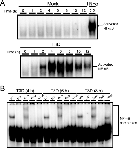
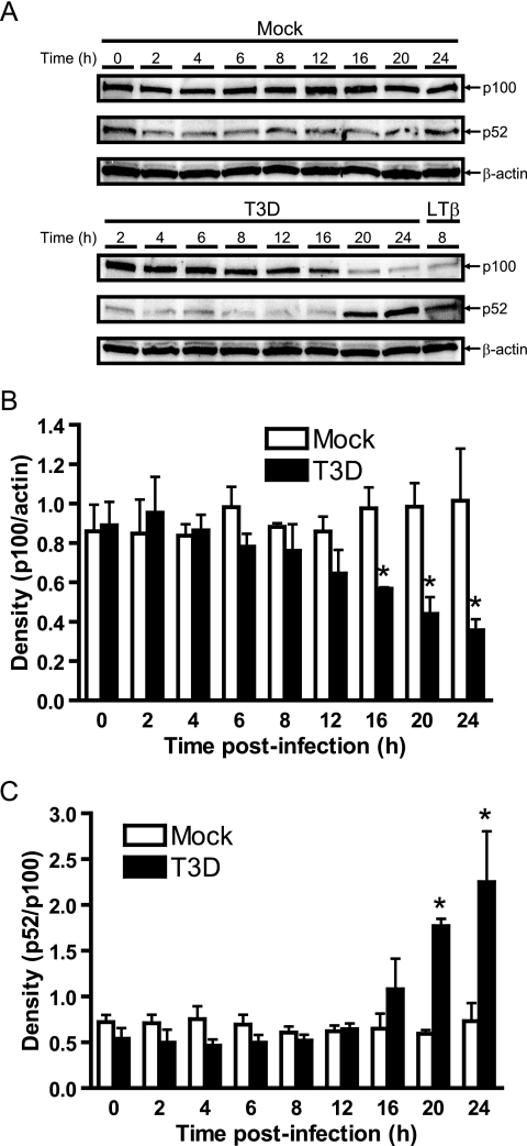
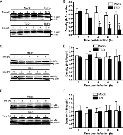
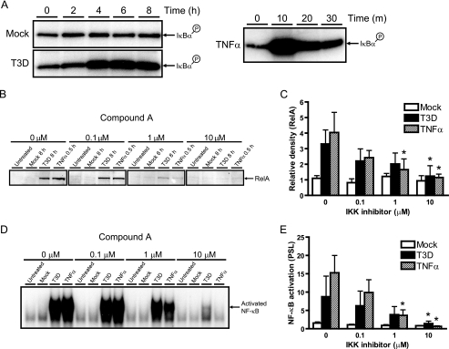
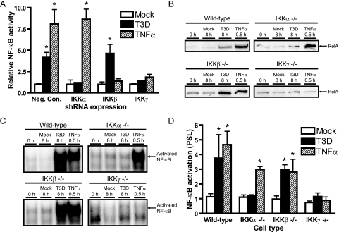
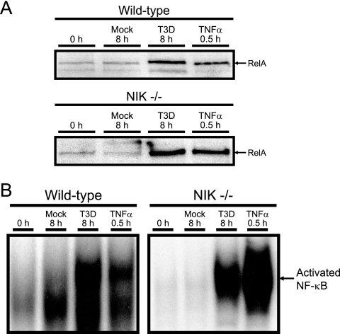
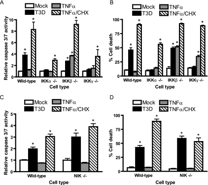
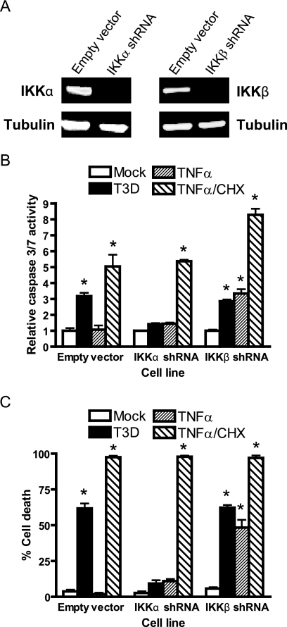
Reoviruses induce apoptosis both in cultured cells and in vivo. Apoptosis plays a major role in the pathogenesis of reovirus encephalitis and myocarditis in infected mice. Reovirus-induced apoptosis is dependent on the activation of transcription factor NF-kappaB and downstream cellular genes. To better understand the mechanism of NF-kappaB activation by reovirus, NF-kappaB signaling intermediates under reovirus control were investigated at the level of Rel, IkappaB, and IkappaB kinase (IKK) proteins. We found that reovirus infection leads initially to nuclear translocation of p50 and RelA, followed by delayed mobilization of c-Rel and p52. This biphasic pattern of Rel protein activation is associated with the degradation of the NF-kappaB inhibitor IkappaBalpha but not the structurally related inhibitors IkappaBbeta or IkappaBepsilon. Using IKK subunit-specific small interfering RNAs and cells deficient in individual IKK subunits, we demonstrate that IKKalpha but not IKKbeta is required for reovirus-induced NF-kappaB activation and apoptosis. Despite the preferential usage of IKKalpha, both NF-kappaB activation and apoptosis were attenuated in cells lacking IKKgamma/Nemo, an essential regulatory subunit of IKKbeta. Moreover, deletion of the gene encoding NF-kappaB-inducing kinase, which is known to modulate IKKalpha function, had no inhibitory effect on either response in reovirus-infected cells. Collectively, these findings indicate a novel pathway of NF-kappaB/Rel activation involving IKKalpha and IKKgamma/Nemo, which together mediate the expression of downstream proapoptotic genes in reovirus-infected cells.


Figures

FIG. 1.
FIG. 1.
Biphasic activation of NF-κB/Rel proteins in reovirus-infected cells. (A) Nuclear extracts were prepared from uninfected HeLa cells (0 h), mock-infected cells (Mock), or cells infected with T3D at an MOI of 100 PFU/cell for the times shown. Cells also were treated with 20 ng/ml of TNF-α for 30 min as a positive control. Extracts were incubated with a radiolabeled NF-κB consensus oligonucleotide, and the resulting protein-oligonucleotide complexes were resolved by polyacrylamide gel electrophoresis, dried, and exposed to film. (B) Nuclear extracts prepared at 4, 6, and 8 h postinfection were incubated with antisera specific for p50, p52, RelA, RelB, or c-Rel prior to the addition of a radiolabeled NF-κB consensus oligonucleotide. NF-κB-containing complexes are indicated.
FIG. 2.
FIG. 2.
Processing of p100 to p52 during reovirus infection. (A) Whole-cell extracts were prepared from uninfected HeLa cells (0 h), mock-infected cells (Mock), or cells infected with reovirus T3D at an MOI of 100 PFU/cell for the times shown. Cells also were treated with 2 μg/ml of an agonistic lymphotoxin-β receptor antiserum for 8 h as a positive control. Extracts were resolved by SDS-PAGE, transferred to nitrocellulose membranes, and immunoblotted by using an antiserum specific for p100/p52. Band intensity was quantified by using the Image J program. The results are expressed as the mean ratio of (B) p100/actin or (C) p52/p100 for three independent experiments. Error bars indicate standard deviations. *, P < 0.05 as determined by Student's t test in comparison to untreated cells (0 h).
FIG. 3.
FIG. 3.
Reovirus infection leads to degradation of IκBα but not IκBβ or IκBɛ. Cytoplasmic extracts were prepared from uninfected HeLa cells (0 h), mock-infected cells (Mock), or cells infected with reovirus T3D at an MOI of 100 PFU/cell for the times shown. Cells also were treated with 20 ng/ml of TNF-α for 10 min as a positive control. Extracts were resolved by SDS-PAGE, transferred to nitrocellulose membranes, and immunoblotted by using antisera specific for (A) IκBα, (C) IκBβ, or (E) IκBɛ. An actin-specific antiserum was used to detect levels of actin as a loading control. Band intensity corresponding to levels of (B) IκBα, (D) IκBβ, and (F) IκBɛ was quantified by using the Image J program. The results are expressed as the mean ratio of IκB/actin for three independent experiments. Error bars indicate standard deviations. *, P < 0.05 as determined by Student's t test in comparison to untreated cells (0 h).
FIG. 4.
FIG. 4.
Involvement of IKKs in reovirus-induced NF-κB activation. (A) Whole-cell extracts were prepared from uninfected HeLa cells (0 h), mock-infected cells (Mock), or cells infected with reovirus T3D at an MOI of 100 PFU/cell for the times shown. Cells also were treated with 20 ng/ml of TNF-α for the times shown as a positive control. The IKK complex was immunoprecipitated by using an IKKγ-specific antiserum prior to incubation with a GST-IκBα substrate in the presence of [γ-32P]ATP. Kinase reactions were resolved by SDS-PAGE, transferred to nitrocellulose, and visualized by autoradiography. (B) HeLa cells were pretreated with IKK inhibitor Compound A for 1 h at the concentrations shown and then uninfected (Untreated), mock infected (Mock), or infected with reovirus T3D at an MOI of 100 PFU/cell for the times shown. Nuclear extracts were resolved by SDS-PAGE, transferred to nitrocellulose, and immunoblotted by using a RelA-specific antiserum. (C) Band intensity was quantified relative to uninfected cells by using the Image J program. The results are expressed as the mean RelA band intensity for three independent experiments. Error bars indicate standard deviations. *, P < 0.05 as determined by Student's t test in comparison to untreated cells (0 μM). (D) Nuclear extracts from the experiment shown in panel B were incubated with a radiolabeled NF-κB consensus oligonucleotide, and the resulting protein-oligonucleotide complexes were resolved by polyacrylamide gel electrophoresis, dried, and exposed to film. (E) Band intensity was quantified by determining PSL units relative to uninfected cells for four independent experiments. Error bars indicate standard deviations. *, P < 0.05 as determined by Student's t test in comparison to untreated cells (0 μM).
FIG. 5.
FIG. 5.
IKKα and IKKγ/Nemo are required for reovirus-induced activation of NF-κB. (A) 293T cells were transfected with plasmids encoding shRNAs specific for IKKα, IKKβ, or IKKγ/Nemo or a negative control shRNA (Neg. Con.). After incubation at 37°C for 48 h, cells were cotransfected with plasmids encoding IKK subunit-specific shRNAs, pNF-κB-Luc, and pRenilla-Luc. Cells were treated with 10 ng/ml of TNF-α and adsorbed with T3D at an MOI of 100 PFU/ml or mock infected prior to assessments of luciferase activity in cell-culture lysates. The results are expressed as the ratio of normalized luciferase activity from infected cell lysates to that from mock-infected lysates for triplicate samples. Error bars indicate standard deviations. (B) Wild-type MEFs or MEFs deficient in IKKα, IKKβ, or IKKγ/Nemo were uninfected (0 h), mock infected (Mock), infected with reovirus T3D at an MOI of 100 PFU/cell for 8 h, or treated with 20 ng/ml of TNF-α for 1 h. Nuclear extracts were resolved by SDS-PAGE, transferred to nitrocellulose, and immunoblotted by using a RelA-specific antiserum. (C) Nuclear extracts from the experiment shown in panel B were incubated with a radiolabeled NF-κB consensus oligonucleotide. Resulting protein-oligonucleotide complexes were resolved by polyacrylamide gel electrophoresis, dried, and exposed to film. NF-κB-containing complexes are indicated. (D) Band intensity was quantified by determining the number of PSL units relative to that in uninfected cells for three independent experiments. Error bars indicate standard deviations. *, P < 0.05 as determined by Student's t test in comparison to mock-treated cells (0 h).
FIG. 6.
FIG. 6.
Reovirus-induced activation of NF-κB in NIK-deficient cells. (A) Wild-type MEFs or NIK-deficient MEFs were uninfected (0 h), mock infected (Mock), infected with reovirus T3D at an MOI of 1,000 PFU/cell for 8 h, or treated with 20 ng/ml of TNF-α for 30 min. Nuclear extracts were resolved by SDS-PAGE, transferred to nitrocellulose, and immunoblotted by using a RelA-specific antiserum. (B) Nuclear extracts from the experiment shown in panel A were incubated with a radiolabeled NF-κB consensus oligonucleotide. The resulting protein-oligonucleotide complexes were resolved by polyacrylamide gel electrophoresis, dried, and exposed to film. NF-κB-containing complexes are indicated.
FIG. 7.
FIG. 7.
IKKα and IKKγ/Nemo are required for reovirus-induced apoptosis. Wild-type MEFs or (A) MEFs deficient in IKKα, IKKβ, IKKγ/Nemo, or (C) NIK were mock infected (Mock), infected with reovirus T3D at an MOI of 1,000 PFU/cell for 24 h (T3D), treated with 10 ng/ml of TNF-α for 12 h (TNFα), or treated with 10 ng/ml of TNF-α and 10 μg/ml of cycloheximide for 12 h (TNFα/CHX). Caspase 3/7 activity was quantified by using a luminescent substrate. The results are expressed as the mean caspase activity relative to that of mock-infected cells for three independent experiments. Wild-type MEFs, (B) IKK-deficient MEFs, or (D) NIK-deficient MEFs were mock infected, infected with reovirus T3D at an MOI of 1,000 PFU/cell for 48 h, treated with 10 ng/ml of TNF-α for 24 h, or treated with 10 ng/ml of TNF-α and 10 μg/ml of cycloheximide for 24 h. Cell viability was quantified by trypan blue exclusion. The results are expressed as the mean percentage of cell death for three independent experiments. Error bars indicate standard deviations. *, P < 0.05 as determined by Student's t test in comparison to mock-infected cells.
FIG. 8.
FIG. 8.
Knockdown of IKKα diminishes apoptosis in response to reovirus. (A) Extracts of HeLa cells transduced with retroviruses containing empty vector or encoding shRNAs specific for either IKKα or IKKβ were resolved by SDS-PAGE, transferred to nitrocellulose, and immunoblotted by using IKKα-, IKKβ-, or tubulin-specific antiserum. Immunoblots were scanned and quantified by using Odyssey software. (B) HeLa cells stably expressing empty vector or shRNAs specific for either IKKα or IKKβ were mock infected (Mock), infected with reovirus T3D at an MOI of 1,000 PFU/cell for 24 h (T3D), treated with 10 ng/ml of TNF-α for 12 h (TNFα), or treated with 10 ng/ml of TNF-α and 10 μg/ml of cycloheximide for 12 h (TNFα/CHX). Caspase 3/7 activity was quantified by using a luminescent substrate. The results are expressed as the mean caspase activity relative to that of mock-infected cells for three independent experiments. (C) HeLa cells stably expressing empty vector or shRNAs specific for either IKKα or IKKβ were mock infected, infected with reovirus T3D at an MOI of 1,000 PFU/cell for 48 h, treated with 10 ng/ml of TNF-α for 24 h, or treated with 10 ng/ml of TNF-α and 10 μg/ml of cycloheximide for 24 h. Cell viability was quantified by trypan blue exclusion. The results are expressed as the mean percentage of cell death for three independent experiments. Error bars indicate standard deviations. *, P < 0.05 as determined by Student's t test in comparison to mock-infected cells.

Similar articles

Cited by

References

    1. Anest, V., J. L. Hanson, P. C. Cogswell, K. A. Steinbrecher, B. D. Strahl, and A. S. Baldwin. 2003. A nucleosomal function for IκB kinase-α in NF-κB-dependent gene expression. Nature 423:659-663. - PubMed
    1. Baeuerle, P., and D. Baltimore. 1989. A 65-kD subunit of active NF-κB is required for inhibition of NF-κB by IκB. Genes Dev. 3:1689-1698. - PubMed
    1. Baeuerle, P., and D. Baltimore. 1988. IκB: a specific inhibitor of the NF-κB transcription factor. Science 242:540-546. - PubMed
    1. Bonizzi, G., and M. Karin. 2004. The two NF-κB activation pathways and their role in innate and adaptive immunity. Trends Immunol. 25:280-288. - PubMed
    1. Bowie, A. G., J. Zhan, and W. L. Marshall. 2004. Viral appropriation of apoptotic and NF-κB signaling pathways. J. Cell. Biochem. 91:1099-1108. - PubMed

Publication types

MeSH terms