. 2006 Nov 15;12(22):6817-25.
doi: 10.1158/1078-0432.CCR-06-1354.

Arsenic trioxide affects signal transducer and activator of transcription proteins through alteration of protein tyrosine kinase phosphorylation

Affiliations

Arsenic trioxide affects signal transducer and activator of transcription proteins through alteration of protein tyrosine kinase phosphorylation

Meir Wetzler et al. Clin Cancer Res. .

Abstract

Purpose: Arsenic trioxide decreases proliferation of acute myeloid leukemia (AML) cells, but its precise mechanism of action is unknown.

Experimental design: We studied the effect of arsenic trioxide on patient samples and the AML cell line HEL, which, like leukemic blasts from 50% of AML cases, has constitutively activated signal transducer and activator of transcription (STAT) proteins.

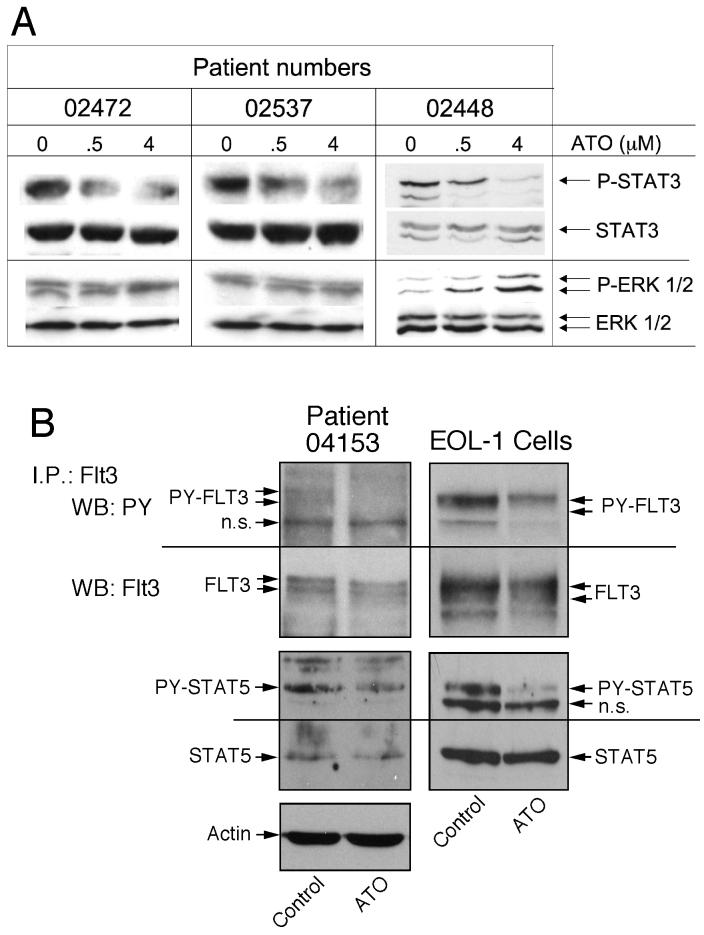
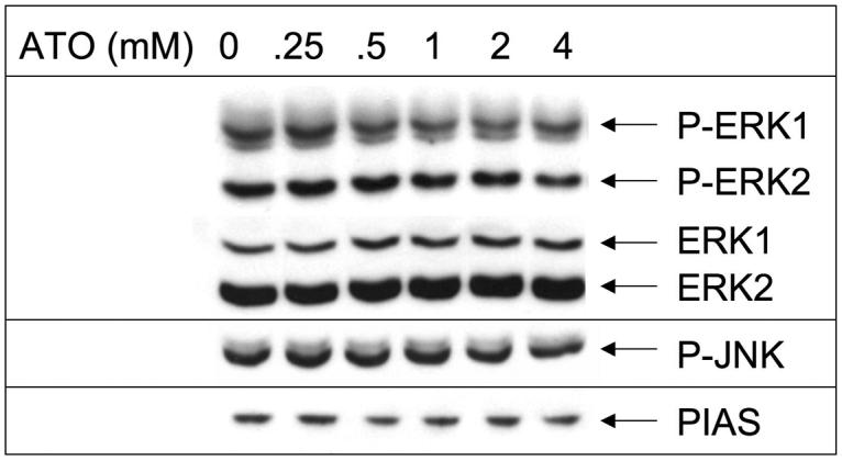
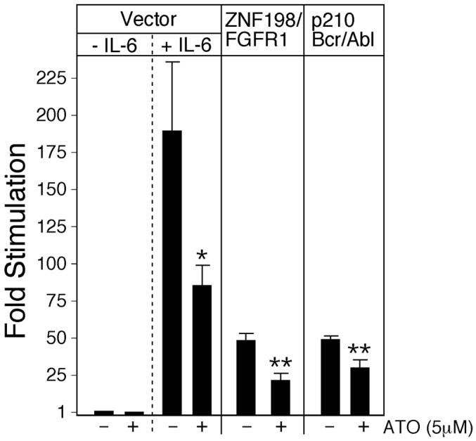
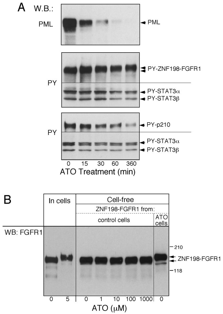
Results: Arsenic trioxide induced mitotic arrest starting at 24 hours and significant cell death at 48 hours. These events were preceded by an arsenic trioxide dose-dependent down-regulation of activated STAT proteins starting at 6 hours. We hypothesized that arsenic trioxide inhibits protein tyrosine kinases (PTK), which, among others, phosphorylate and activate STATs. We therefore studied arsenic trioxide effects on Janus kinases and on three oncogenic PTKs that are known to activate STATs [FLT3, ZNF198/fibroblast growth factor receptor 1 (FGFR1), and BCR/ABL]. Arsenic trioxide reduced STAT3 activation by Janus kinases, altered phosphorylation and electrophoretic mobility of ZNF198/fibroblast growth factor receptor 1, reduced kinase protein level, and decreased STAT3 protein phosphorylation. Arsenic trioxide also reduced the phosphorylation of BCR/ABL and FLT3 with corresponding decreased STAT5 phosphorylation.

Conclusions: These results suggest a selective activity of arsenic trioxide on PTKs and will assist in developing clinical trials in AML.


Figures

Figure 1
Figure 1
ATO down regulates STAT activity in AML blasts. Cells were exposed to ATO at 2 concentrations for 6 hours. A, samples from 3 AML patients with no known oncogenic PTK activity. ATO induced minimal phosphorylation of ERK 1/2 in the sample from patient 02448 but not in the other 2 patient samples. B, left panel, a sample from a patient with FLT3-ITD demonstrating down-regulation of FLT3-ITD and phosphorylated STAT5 by 4 μM ATO. B, right panel, control EOL-1 cells with constitutively active FLT3.
Figure 2
Figure 2
ATO decreases survival of HEL cells through an arrest at the G2/M stage of the cell cycle. A, Hill model curve demonstrating the effect of ATO at different concentrations on total cell count at 48 hours. B, mitotic arrest (right panel) induced by ATO at 24 hours as compare to control (left panel) cells (May-Grünwald stain, magnification x40). C and D, cell cycle analysis of control cells demonstrating 9% cells in G2/M (C) and cells treated with 4 μM ATO for 24 hours demonstrating 20% cells in G2/M (D).
Figure 3
Figure 3
ATO downregulates constitutive STAT1, STAT3 and STAT5 activities in HEL cells. Cells were exposed to ATO at different concentrations for 6 hours. A, western blotting and EMSA (demonstrating the SIE-binding activity of STAT1 and STAT3) analyses in the upper panel and western blotting demonstrating STAT5 activity in the lower panel. B, C and D, Hill model analyses of cumulative western blotting data for phosphorylated STAT1 (panel B), STAT3 (panel C) and STAT5 (panel D) from multiple experiments.
Figure 4
Figure 4
ATO does not affect phosphorylation of ERK 1/2 or JNK and does not modify PIAS3 levels. Western blot analysis following 6 hours of incubation with ATO.
Figure 5
Figure 5
ATO modulates oncogenic and non-oncogenic PTKs. 293 cells in 6-well plates were transfected with the expression vectors for ZNF198/FGFR1 (A), GFP-tagged-ZNF198/FGFR1 (B), BCR/FGFR1 (C), BCR/ABL (C), JAK1 (D), or JAK2 (D) (4μg DNA/well). The cells were treated 24 hours later with the indicated concentration of ATO (A and B) 5 μM (C and D) for 6 hours. Cells were extracted and aliquots of each extract were electrophoresed on separate gels. The blots with transferred proteins were reacted with Ab as shown on the left. The positions of the transfected proteins are indicated on the right. Panel B, Fluorescent images of the transfected cells were taken at 40x magnification. WB, western blotting.
Figure 6
Figure 6
ATO decreased transcription of the STAT3-responsive gene, HPX-IL-6RE. HepG2 cells were transfected with p(5xHPX-IL-6RE)-CAT (15 μg/ml) together with an empty expression vector, or an expression vector for ZNF198/FGFR1 or BCR/ABL, as indicated at the top of the panels. The subcultures of the transfected cells were treated for 24 hours with medium alone or with ATO. The panel represents the PhosphorImager quantification (mean ± SD) of the fold stimulation values relative to the untreated vector control culture in 3 independently performed experiments. *, p<0.05; **, p<0.005.
Figure 7
Figure 7
ATO mediates a modification of ZNF198/FGFR1 distinct from PML and BCR/ABL. A: ATO mediated degradation of PML is kinetically distinct from change in phosphorylation of BCR/ABL, ZNF198/FGFR1 or STAT3. 293 cells transfected with PML, BCR/ABL, ZNF198/FGFR1 and STAT3 were exposed to 5 μM ATO and harvested at different time points. Total cell extracts were subjected to immunoblotting with the corresponding Ab. B: ATO requires the cell environment to exert its effect on ZNF198/FGFR1. 293 cells were transfected with ZNF198/FGFR1 and subcultures were treated for 30 min with medium alone (control cells) or medium with 5 μM ATO (ATO cells). Cytosolic extracts were prepared from each culture. Aliquots from the control cells were incubated with ATO at the indicated concentrations for 30 min at 370 followed by Western blot (WB) analysis with the anti-FGFR1 antibody. An aliquot of extract from ATO cells was incubated for 30 min without ATO and served as indicator for potential, ATO-independent modifications during the cell-free reaction.

References

    1. Benekli M, Baer MR, Baumann H, Wetzler M. Signal transducer and activator of transcription proteins in leukemias. Blood. 2003;101:2940–2954. - PubMed
    1. Xia Z, Baer MR, Block AW, Baumann H, Wetzler M. Expression of signal transducers and activators of transcription proteins in acute myeloid leukemia blasts. Cancer Res. 1998;58:3173–3180. - PubMed
    1. Benekli M, Xia Z, Donohue KA, Ford LA, Pixley LA, Baer MR, Baumann H, Wetzler M. Constitutive activity of signal transducer and activator of transcription 3 protein in acute myeloid leukemia blasts is associated with short disease-free survival. Blood. 2002;99:252–257. - PubMed
    1. Shen ZX, Chen GQ, Ni JH, Li XS, Xiong SM, Qiu QY, Zhu J, Tang W, Sun GL, Yang KQ, Chen Y, Zhou L, Fang ZW, Wang YT, Ma J, Zhang P, Zhang TD, Chen SJ, Chen Z, Wang ZY. Use of arsenic trioxide (As2O3) in the treatment of acute promyelocytic leukemia (APL): II. Clinical efficacy and pharmacokinetics in relapsed patients. Blood. 1997;89:3354–3360. - PubMed
    1. Bachleitner-Hofmann T, Gisslinger B, Grumbeck E, Gisslinger H. Arsenic trioxide and ascorbic acid: synergy with potential implications for the treatment of acute myeloid leukaemia? Br J Haematol. 2001;112:783–786. - PubMed

Publication types

MeSH terms