Comparative Study
. 2006 Nov 22;26(47):12274-82.
doi: 10.1523/JNEUROSCI.2599-06.2006.

Differential expression of I(A) channel subunits Kv4.2 and Kv4.3 in mouse visual cortical neurons and synapses

Affiliations
Comparative Study

Differential expression of I(A) channel subunits Kv4.2 and Kv4.3 in mouse visual cortical neurons and synapses

Andreas Burkhalter et al. J Neurosci. .

Abstract

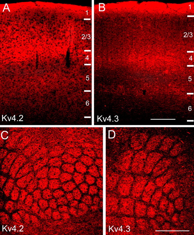
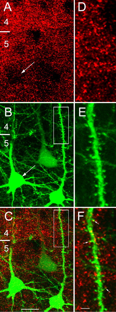
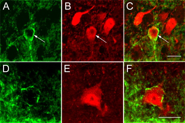
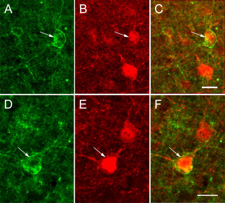
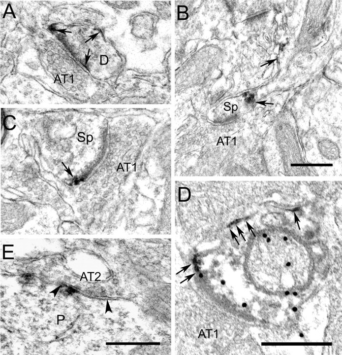
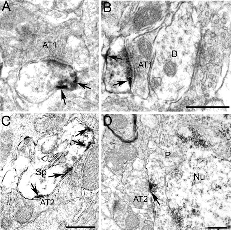
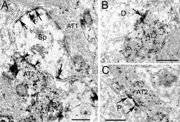
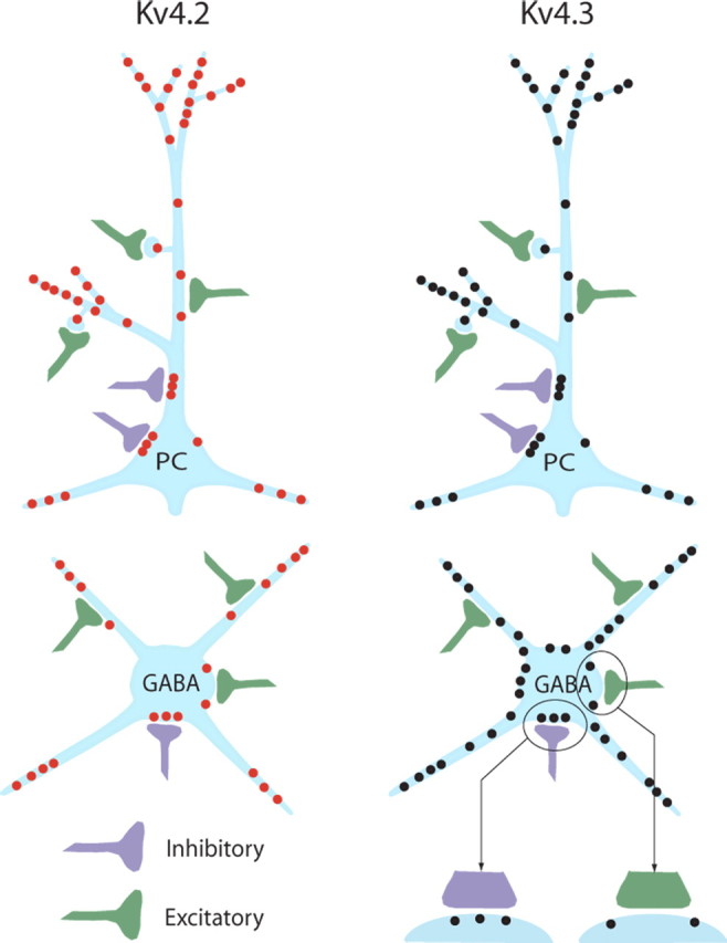
In cortical neurons, pore-forming alpha-subunits of the Kv4 subfamily underlie the fast transient outward K+ current (I(A)). Considerable evidence has accumulated demonstrating specific roles for I(A) channels in the generation of individual action potentials and in the regulation of repetitive firing. Although I(A) channels are thought to play a role in synaptic processing, little is known about the cell type- and synapse-specific distribution of these channels in cortical circuits. Here, we used immunolabeling with specific antibodies against Kv4.2 and Kv4.3, in combination with GABA immunogold staining, to determine the cellular, subcellular, and synaptic localization of Kv4 channels in the primary visual cortex of mice, in which subsets of pyramidal cells express yellow fluorescent protein. The results show that both Kv4.2 and Kv4.3 are concentrated in layer 1, the bottom of layer 2/3, and in layers 4 and 5/6. In all layers, clusters of Kv4.2 and Kv4.3 immunoreactivity are evident in the membranes of the somata, dendrites, and spines of pyramidal cells and GABAergic interneurons. Electron microscopic analyses revealed that Kv4.2 and Kv4.3 clusters in pyramidal cells and interneurons are excluded from putative excitatory synapses, whereas postsynaptic membranes at GABAergic synapses often contain Kv4.2 and Kv4.3. The presence of Kv4 channels at GABAergic synapses would be expected to weaken inhibition during dendritic depolarization by backpropagating action potentials. The extrasynaptic localization of Kv4 channels near excitatory synapses, in contrast, should stabilize synaptic excitation during dendritic depolarization. Thus, the synapse-specific distribution of Kv4 channels functions to optimize dendritic excitation and the association between presynaptic and postsynaptic activity.


Figures

Figure 1.
Figure 1.
Expression levels of Kv4.2 and Kv4.3 in mouse primary visual and primary somatosensory cortex are layer specific. A, B, Immunofluorescence images of Kv4.2 and Kv4.3 expression in coronal sections through mouse primary visual cortex. Staining is densest in layers 1 and 4 and the layer 5/6 border. C, D, Kv4.2 and Kv4.3 immunofluorescence in tangential sections through mouse primary somatosensory cortex. Labeling is most intense in layer 4, in the center of barrels. Expression in septa between barrels is sparse. Scale bars: A, B, 0.2 mm; C, D, 1 mm.
Figure 2.
Figure 2.
Kv4.2 clusters are expressed in pyramidal cell somata, dendrites, and spines. A, Confocal microscopic images of punctate Kv4.2 immunolabeling (red) in layers 4 and 5 of mouse primary visual cortex. The distribution of Kv4.2 clusters is non-uniform, showing a higher density over the neuropil than the pyramidal cell soma in the same focal plane (arrows; A, B). B, C, Kv4.2 clusters (red, yellow) are associated with YFP-labeled (green) pyramidal cell somata and with basal and apical dendrites. DF, High-magnification images of the boxed apical dendrite of a YFP-labeled layer 5 pyramidal neuron (B, C), showing close association of Kv4.2-labeled clusters (red and yellow puncta marked with arrows) with dendritic shaft and spines. The numbers indicate layers. Scale bars: AC, 10 μm; DF, 2 μm.
Figure 3.
Figure 3.
Kv4.2 expression in GABAergic neurons of mouse primary visual cortex. Confocal microscopic images of immunolabeled GABAergic neurons (red) in layer 2/3 (AC) and layer 5 (DF). Kv4.2 (green) is coexpressed in somata and dendrites of a subset of GABAergic neurons. Arrows in AC mark Kv4.2-expressing GABAergic neuron. Scale bars, 10 μm.
Figure 4.
Figure 4.
Confocal microscopic images of Kv4.3 immunolabeling in pyramidal neurons of mouse primary visual cortex. AC, YFP-labeled pyramidal neuron with a soma in layer 5 and sparsely spiny apical dendrite in layers 4 and 2/3 that are decorated with Kv4.3 clusters (red and yellow puncta marked with arrows). The arrowhead indicates a spine, the neck of which contains a Kv4.3-positive cluster. DF, Kv4.3 expression (red; D) in layer 2/3 pyramidal neurons that are stained with an antibody against neurofilament (green; E). Kv4.3 clusters (arrows) that are associated with somata and dendrites appear yellow (F). The numbers indicate layers. Scale bars: AC, 10 μm; DF, 20 μm.
Figure 5.
Figure 5.
Confocal microscopic images of Kv4.3 immunolabeling in GABAergic neurons of mouse primary visual cortex. AC, Coexpression (arrows) of Kv4.3 (green; A) and GABA (red; B) in a subset of layer 2/3 nonpyramidal cells (C) in which Kv4.3 labeling extends into distal branches of the dendritic tree. DF, Coexpression (arrows) of Kv4.3 (green; D) and GABA (red; E) in a subset of layer 5 nonpyramidal neurons (F). Scale bars, 10 μm.
Figure 6.
Figure 6.
Electron micrographs showing that Kv4.2 is differentially expressed at putative inhibitory and excitatory synapses in mouse primary visual cortex. A, Extrasynaptic localization of Kv4.2 immunoperoxidase labeling (arrows) at an asymmetric synapse onto a thin dendrite (D). The dark immunoperoxidase reaction product is easily distinguished from the much lighter postsynaptic density. B, Kv4.2 immunoperoxidase at the base and neck of spine. Kv4.2 is absent from the asymmetric synapse. C, Extrasynaptic localization of Kv4.2 at an asymmetric synapse onto spine. D, Extrasynaptic localization of Kv4.2 (arrows) at an asymmetric synapse onto GABA immunogold (black dots)-labeled dendrite. E, Synaptic localization of Kv4.2 at a symmetric synapse (AT2) onto a pyramidal cell soma (P). Notice that Kv4.2 is contained in the postsynaptic membrane associated with the synaptic cleft, the margins of which are indicated by arrowheads. Scale bars, 0.5 μm. AT1, Asymmetric synapse; Sp, spine.
Figure 7.
Figure 7.
Electron micrographs showing that Kv4.3 is differentially expressed at putative inhibitory and excitatory synapses in mouse primary visual cortex. A, B, Kv4.3 immunoperoxidase labeling (dark reaction product marked by arrows) in thin dendrites is excluded from asymmetric synapses (AT1). The thick dendrite (D) does not express Kv4.3. C, D, Kv4.3 immunoperoxidase labeling (arrows) in postsynaptic membranes directly across Gray type 2 axon terminals (AT2) that contain flattened vesicles and form symmetric, putative inhibitory synapses with spine (Sp) and perikaryon (P) of a nonpyramidal cell [identified by infoldings of nuclear (Nu) membrane]. Scale bars, 0.5 μm.
Figure 8.
Figure 8.
Electron micrographs showing differential localization of Kv4.2 and Kv4.3 at GABAergic and non-GABAergic synapses in mouse primary visual cortex. A, Kv4.2 immunoperoxidase (arrows and arrowheads) expression in a GABA immunogold (black dots)-labeled section. Notice that Kv4.2 (arrows) is expressed in the spine (Sp) membrane but is excluded from the asymmetric synapse formed by a non-GABAergic Gray type 1 axon terminal (AT1). In contrast, Kv4.2 is expressed at a symmetric synapse formed by GABA immunogold-labeled Gray type 2 axon terminal. B, C, Postsynaptic localization of Kv4.3 immunoperoxidase labeling (arrows) at symmetric synapses formed by GABAergic (black dots) Gray type 2 axon terminals onto dendrite (D) and perikaryon (P). Scale bars, 0.5 μm.
Figure 9.
Figure 9.
Schematic representation of Kv4.2 (red dots) and Kv4.3 (black dots) subunit distribution in neocortical pyramidal cells (top) and GABAergic interneurons (bottom). A close-up at the bottom right applies to both Kv4.2 and Kv4.3 and shows that Kv4 channels are excluded from excitatory synapses (green) but are present at GABAergic inhibitory synapses (purple). PC, Pyramidal cell.

References

    1. Alonso G, Widmer H. Clustering of Kv4.2 potassium channels in postsynaptic membrane of rat supraoptic neurons: an ultrastructural study. Neuroscience. 1997;77:617–621. - PubMed
    1. Antonini A, Fagolini M, Stryker MP. Anatomical correlates of functional plasticity in mouse visual cortex. J Neurosci. 1999;19:4388–4406. - PMC - PubMed
    1. Cai X, Liang CW, Muralidharan S, Kao JPY, Tang C-M, Thompson SM. Unique roles of SK and Kv4.2 potassium channels in dendritic integration. Neuron. 2004;44:351–364. - PubMed
    1. Chu P-J, Rivera JF, Arnold DB. A role for Kif17 in transport of Kv4.2. J Biol Chem. 2006;281:365–373. - PubMed
    1. Chu Z, Galarreta M, Hestrin S. Synaptic interactions of late-spiking neocortical neurons in layer 1. J Neurosci. 2003;23:96–102. - PMC - PubMed

Publication types