

Eye movements of patients with tunnel vision while walking
- PMID: 17122116
- PMCID: PMC1752198
- DOI: 10.1167/iovs.05-1043
Eye movements of patients with tunnel vision while walking
Abstract
Purpose: To determine how severe peripheral field loss (PFL) affects the dispersion of eye movements relative to the head in patients walking in real environments. This information should help to define the visual field and clearance requirements for head-mounted mobility visual aids.
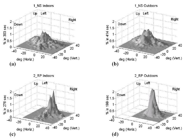
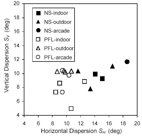
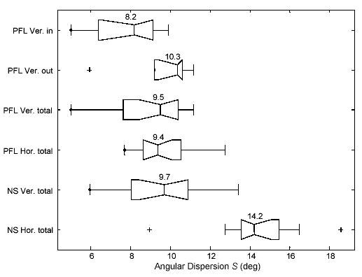
Methods: Eye positions relative to the head were recorded in five patients with retinitis pigmentosa who had less than 15 degrees of visual field and in three normally sighted people, each walking in varied environments for more than 30 minutes. The eye-position recorder was made portable by modifying a head-mounted system (ISCAN, Burlington, MA). Custom data processing was implemented, to reject unreliable data. Sample standard deviations of eye position (dispersion) were compared across subject groups and environments.
Results: The patients with PFL exhibited narrower horizontal eye-position dispersions than did the normally sighted subjects (9.4 degrees vs. 14.2 degrees , P < 0.0001), and the vertical dispersions of patients with PFL were smaller when they were walking indoors than when walking outdoors (8.2 degrees vs. 10.3 degrees ; P = 0.048).
Conclusions: When walking, the patients with PFL did not increase their scanning eye movements to compensate for missing peripheral vision information. Their horizontal scanning was actually reduced, possibly because of lack of peripheral stimulation. The results suggest that a field of view as wide as 40 degrees may be needed for closed (immersive) head-mounted mobility aids, whereas a much narrower display, perhaps as narrow as 20 degrees , may be sufficient with an open design.
Figures

References
-
- Robinson B, Acorn CJ, Millar CC, Lyle WM. The prevalence of selected ocular diseases and conditions. Optom Vis Sci. 1997;74:79–91. - PubMed
-
- Turano KA, Rubin GS, Quigley HA. Mobility performance in glaucoma. Invest Ophthalmol Vis Sci. 1999;40:2803–2809. - PubMed
-
- Turano KA, Geruschat DR, Stahl JW, Massof RW. Perceived visual ability for independent mobility in persons with retinitis pigmentosa. Invest Ophthalmol Vis Sci. 1999;40:865–877. - PubMed
-
- Pagon RA. Retinitis pigmentosa. Surv Ophthalmol. 1988;33:137–177. - PubMed
-
- Dickinson C. Oxford: Butterworth-Heinemann; 1998. Low Vision: Principles and Practice.
Publication types
MeSH terms
Grants and funding
LinkOut - more resources
Full Text Sources
Other Literature Sources
Medical
