. 2007 Feb;18(2):475-86.
doi: 10.1091/mbc.e06-06-0546. Epub 2006 Nov 22.

The Tim9p/10p and Tim8p/13p complexes bind to specific sites on Tim23p during mitochondrial protein import

Affiliations

The Tim9p/10p and Tim8p/13p complexes bind to specific sites on Tim23p during mitochondrial protein import

Alison J Davis et al. Mol Biol Cell. 2007 Feb.

Abstract

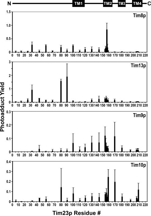
The import of polytopic membrane proteins into the mitochondrial inner membrane (IM) is facilitated by Tim9p/Tim10p and Tim8p/Tim13p protein complexes in the intermembrane space (IMS). These complexes are proposed to act as chaperones by transporting the hydrophobic IM proteins through the aqueous IMS and preventing their aggregation. To examine the nature of this interaction, Tim23p molecules containing a single photoreactive cross-linking probe were imported into mitochondria in the absence of an IM potential where they associated with small Tim complexes in the IMS. On photolysis and immunoprecipitation, a probe located at a particular Tim23p site (27 different locations were examined) was found to react covalently with, in most cases, only one of the small Tim proteins. Tim8p, Tim9p, Tim10p, and Tim13p were therefore positioned adjacent to specific sites in the Tim23p substrate before its integration into the IM. This specificity of binding to Tim23p strongly suggests that small Tim proteins do not function solely as general chaperones by minimizing the exposure of nonpolar Tim23p surfaces to the aqueous medium, but may also align a folded Tim23p substrate in the proper orientation for delivery and integration into the IM at the TIM22 translocon.


Figures

Figure 1.
Figure 1.
Photocross-linking of substrates of the TIM22 pathway to mitochondrial import components. (A) Photoadducts vary with stage of import. Tim23p and Aac2p were translated separately in the presence of [35S]Met and either Lys-tRNALys (−ANB) or εANB-Lys-tRNALys (+ANB). Translation products were added to mitochondria containing (+ΔΨ) or lacking (−ΔΨ) an inner membrane potential as described in Materials and Methods, and samples were then photolyzed and sedimented to isolate the mitochondria. Mitochondrial proteins were separated by SDS-PAGE, and radiolabeled proteins were visualized by phosphorimaging. Lanes 1, 2, 7, and 8 contain 1/10 of the volume of the translation mix that was added to each import reaction. Arrowheads identify photoadduct bands. p, Tim23p or Aac2p. (B) Tiny Tim photoadducts. Import intermediates containing Tim23p or Aac2p were prepared as in A with εANB-Lys-tRNALys and −ΔΨ mitochondria, photolyzed, and split into five portions before mitochondria were pelleted. One aliquot (5 μg) was resuspended directly in sample buffer (XL tot), whereas the other aliquots (50 μg) were immunoprecipitated with antibodies specific for Tim8p, Tim9p, Tim10p, or Tim13p as indicated. Other labels are the same as in A. (C) IMS complexes of Tim23p with small Tim proteins are productive intermediates. Tim23p import intermediates were prepared as in A with εANB-Lys-tRNALys and −ΔΨ mitochondria, sedimented, and resuspended either in import buffer containing CCCP (−ΔΨ) or in import buffer containing 2 mM malate, 2 mM pyruvate, and DTT as indicated (+ΔΨ) to restore the IM potential. After photolysis, samples were immunoprecipitated using antibodies specific for Tim13p (lanes 1, 4, 7, and 10), Tim50p (lanes 2, 5, 8, and 11), and the unrelated HA epitope (lanes 3, 6, 9, and 12).
Figure 2.
Figure 2.
Tim23p derivatives containing a single εANB-Lys are import competent and form stage-specific photoadducts. (A) Schematic representation of the integrated Tim23p protein and the locations of photoreactive probes. The numbered rectangles indicate the four hydrophobic regions proposed to be membrane-spanning segments of Tim23p. Small numbers along the length of the Tim23p sequence denote the amino acid positions at which an amber codon was substituted for the natural codon. (B) Suppressor tRNAs translate amber codons located far into the coding sequence. Tim23p containing amber codons at amino acid positions 111, 156, 169, 181, 195, or 206 were translated in vitro with [35S]Met and in the presence or absence of Lys-tRNAamb. The full-length (suppressed) nascent chains are ∼27 kDa and are marked by an arrowhead; the nonsuppressed nascent chains terminated at the amber codon are indicated by the bracket. (C) Tim23p containing an εANB-Lys probe is imported into mitochondria and integrated into the IM. Wild-type Tim23p, TAG-131, and TAG-156 were translated in the presence of εANB-Lys -tRNAamb and then exposed to 10 mM DTT to chemically inactivate the azido group. Each precursor was imported into isolated mitochondria, and the samples were split into three 25-μg aliquots. One aliquot was not treated further (lanes 2, 7, and 12), one aliquot was treated with 50 μg/ml proteinase K on ice for 20 min (lanes 3, 8, and 13), and the mitochondria in the third aliquot were converted to mitoplasts by osmotic shock and then treated with proteinase K (lanes 4, 9, and 14). Each precursor was also imported into mitochondria (25 μg) lacking a membrane potential (−ΔΨ), converted to mitoplasts, and treated with proteinase K (lanes 5, 10, and 15). In all samples, mitochondria or mitoplasts were pelleted before analysis by SDS-PAGE and phosphorimaging. Arrowheads indicate the protease-protected fragment of Tim23p in mitoplasts, indicative of successful integration of Tim23p into the IM. (D) Photocross-linking to Tim23p varies with probe location and the stage of integration. Tim23p TAG-25, TAG-32, and TAG-66 were translated with either εANB-Lys-tRNAamb or Lys-tRNAamb (+ or −ANB), imported into mitochondria in the presence or absence of a membrane potential (+/−ΔΨ), and then photocross-linked and analyzed as above. Prominent photoadducts are indicated by arrowheads (lanes 4 and 8) and an asterisk (lane 10).
Figure 3.
Figure 3.
Site-specific photocross-linking of Tim23p to Tim8p and Tim9p in the IMS. TIM23 constructs with single amber codons located at 27 different positions (aa#) were transcribed and translated in the presence of [35S]Met and εANB-Lys-tRNAamb, imported into de-energized mitochondria, and photolyzed. Samples were split into two aliquots and pelleted: one aliquot (5 μg) was resuspended in sample buffer and the other (50 μg) was immunoprecipitated as described in Materials and Methods, first with antisera to Tim8p (αTim8p) and subsequently with antisera to Tim9p (αTim9p) before analysis by SDS-PAGE and phosphorimaging. (A) Total radioactive species; (B) photoadducts immunoprecipitated with αTim8p or αTim9p. 23p, Tim23p; nonsup., nonsuppressed Tim23p (these proteins are not visible in most lanes because the earlier location of the amber stop codons created shorter terminated polypeptides that ran off the gel).
Figure 4.
Figure 4.
Site-specific photocross-linking of Tim23p to Tim10p and Tim13p in the IMS. Experiments were performed exactly as in Figure 3, except that samples were first immunoprecipitated with αTim10p and then with αTim13p. (A) Total radioactive species; (B) photoadducts immunoprecipitated with αTim10p or αTim13p.
Figure 5.
Figure 5.
Quantification of photoadduct formation. The photoadduct yield (the ratio of Tim23p photoadduct to unreacted Tim23p imported into mitochondria) was determined after immunoprecipitation with αTim8p-, αTim9p-, αTim10p-, or αTim13p-specific antiserum as described in Materials and Methods. Bars show the averaged data and SDs from three independent experiments. Probe locations in the Tim23p sequence are indicated on the x-axis, and the putative locations of the TMSs in Tim23p are indicated in the linear representation of the Tim23p sequence at the top of the figure.
Figure 6.
Figure 6.
Photocross-linking of sites in Tim23p TMS2 to the IMS proteins. The data shown in Figure 5 have been normalized as described in the text to depict the relative photocross-linking efficiencies of probes positioned at residues 153-158 in Tim23p to each of the small Tim proteins.
Figure 7.
Figure 7.
Site-specific photocross-linking of Tim23Cp to Tim9p or Tim10p. Experiments were performed exactly as in Figure 3, except that samples were first immunoprecipitated with αTim9p and then with αTim10p. (A) Total radioactive species; (B) photoadducts immunoprecipitated with αTim 9p or αTim10p; (C) the photoadduct yield was determined after immunoprecipitation with αTim9p or αTim10p as in Figure 5.

References

    1. Adam A., Endres M., Sirrenberg C., Lottspeich F., Neupert W., Brunner M. Tim9, a new component of the TIM22Á54 translocase in mitochondria. EMBO J. 1999;18:313–319. - PMC - PubMed
    1. Attardi G., Schatz G. Biogenesis of mitochondria. Annu. Rev. Cell Biol. 1988;4:289–333. - PubMed
    1. Bauer M. F., Sirrenberg C., Neupert W., Brunner M. Role of Tim23 as voltage sensor and presequence receptor in protein import into mitochondria. Cell. 1996;87:33–41. - PubMed
    1. Curran S. P., Leuenberger D., Oppliger W., Koehler C. M. The Tim9p-Tim10p complex binds to the transmembrane domains of the ADP/ATP carrier. EMBO J. 2002a;21:942–953. - PMC - PubMed
    1. Curran S. P., Leuenberger D., Schmidt E., Koehler C. M. The role of the Tim8p-Tim13p complex in a conserved import pathway for mitochondrial polytopic inner membrane proteins. J. Cell Biol. 2002b;158:1017–1027. - PMC - PubMed

Publication types

MeSH terms

Substances

LinkOut - more resources