. 2006 Dec;244(6):986-92; discussion 992-3.
doi: 10.1097/01.sla.0000247058.43243.7b.

Tumor infiltrating T lymphocytes in colorectal cancer: Tumor-selective activation and cytotoxic activity in situ

Affiliations

Tumor infiltrating T lymphocytes in colorectal cancer: Tumor-selective activation and cytotoxic activity in situ

Moritz Koch et al. Ann Surg. 2006 Dec.

Abstract

Objective: To examine whether tumor-selective infiltration, activation, and cytotoxic activity of tumor infiltrating T lymphocytes (TIL) can be demonstrated in situ in colorectal cancer samples.

Summary background data: Recent studies indicated a correlation between the presence of TIL and an improved prognosis in colorectal cancer. However, tumor-selective activation and cytotoxic activity of CD8 TIL in situ in colorectal cancer patients have not yet been examined.

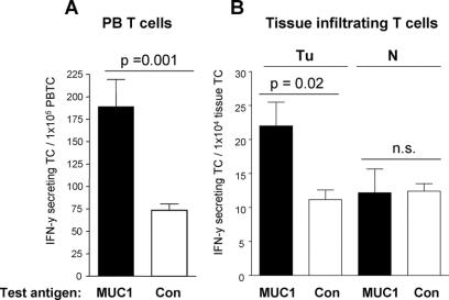
Methods: Tumor samples from 49 patients and corresponding normal mucosa samples from 23 patients with colorectal cancer (UICC stages II-IV) were examined for TIL. Two-color fluorescence immunohistochemistry and multicolor flowcytometric (FACS) analysis were used for quantification of CD8 T cells and measurement of their activation status (CD69-expression) and cytotoxic activity (CD107a-expression) in situ. Presence of tumor antigen-reactive T cells in tumor, blood, and bone marrow was evaluated by IFN-gamma Elispot analysis.

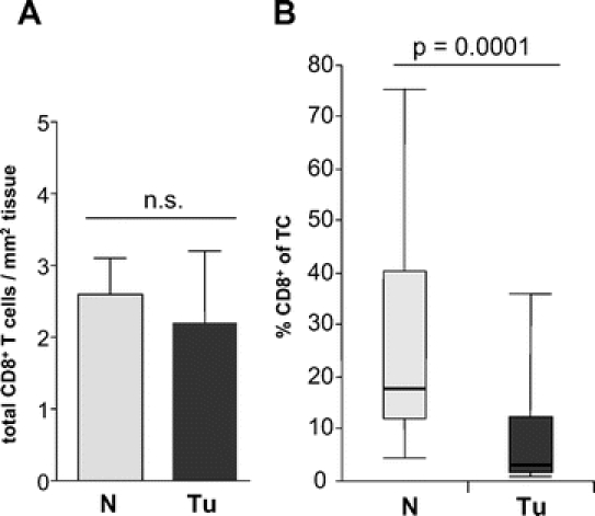
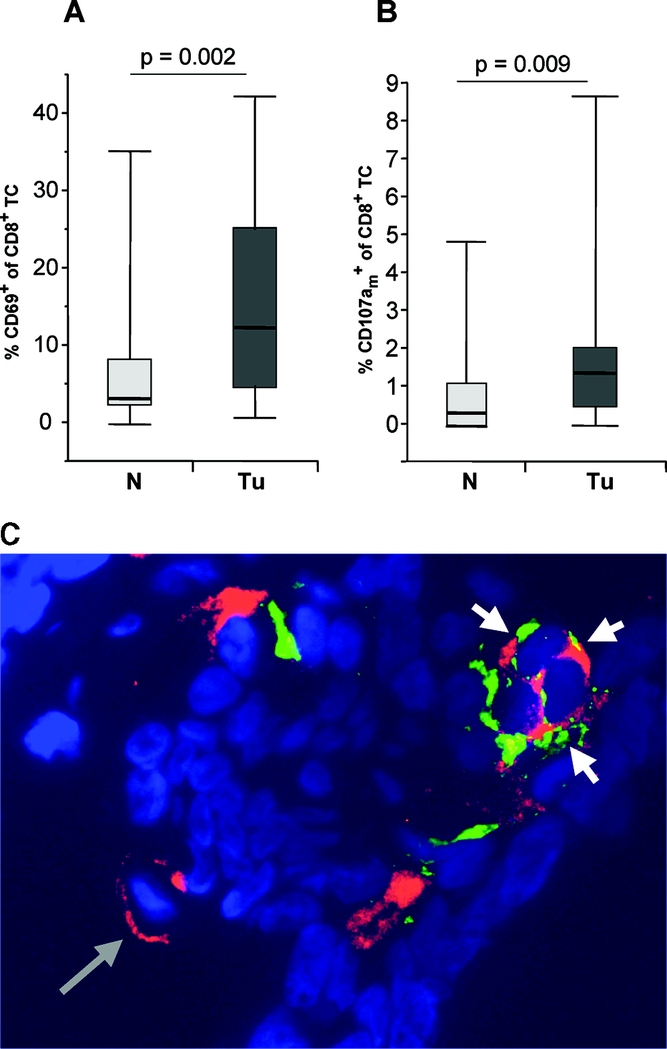
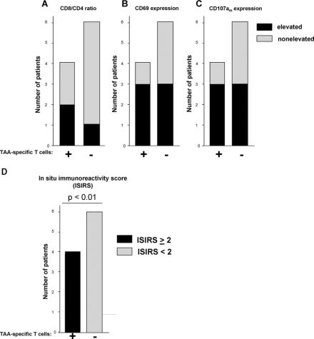
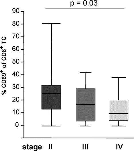
Results: While absolute numbers of CD8 T cells were similar, CD4 T helper cells were significantly increased in tumor tissue compared with normal mucosa. There was a significantly higher proportion of activated and cytotoxically active CD8 TIL in colorectal cancer compared with normal mucosa. Increased activation, cytotoxic activity, and functional reactivity of TIL were correlated with the presence of functional tumor antigen-reactive T cells in the blood and bone marrow. The proportion of activated TIL decreased significantly with higher tumor stage.

Conclusions: Tumor-selective activation and cytotoxic activity of CD8 TIL and tumor-selective migration of CD4 T helper cells were demonstrated in colorectal cancer for the first time. Our data support the immunogenicity of colorectal cancer and suggest clinical significance of tumor-specific immune responses.


Figures

None
FIGURE 1. CD8+ T cell infiltration in 23 colorectal carcinomas (Tu) and 23 normal mucosa samples (N). A, Mean ± SEM CD8+ T cell counts per mm2 tumor tissue or normal mucosa. n.s., no significant difference between groups. B, Box and whisker plots of the percentage of CD8+ T cells among total (CD4+ and CD8+) T cells (TC) as determined by 2-color fluorescence immunohistology. Upper and lower endpoints of the whiskers represent the maximum and minimum percentages. The upper and lower edges of the boxes represent the 75th and 25th percentiles. The line inside the box represents the median.
None
FIGURE 2. Selective activation and cytotoxic activity of tumor infiltrating CD8 T cells. Box and whisker plots of the percentage of the proportions of CD69+ (A) and CD107am+ (B) cells among CD8+ T cells analyzed in 23 colorectal carcinomas (Tu) and 23 normal mucosa samples (N) of corresponding patients using 2-color fluorescence immunohistology (A) or multicolor flow cytometry (B). Upper and lower end points of the whiskers represent the maximum and minimum percentages. The upper and lower edges of the boxes represent the 75th and 25th percentiles. The line inside the box represents the median. (a similar difference was detected by 2-color immunohistology of the same tissues; data not shown) C, One representative histologic tumor section stained for CD8 (red) and CD107a (green). Nuclei are counterstained in blue color. A single CD8+ T cell is depicted by a long gray arrow. Cytotoxically active CD8+ T cells with membranous expression of CD107am are depicted by short white arrows. Original magnification ×200.
None
FIGURE 3. MUC1-reactive T cells in the blood and tumor tissue of colorectal carcinoma patients; results of one representative patient. Numbers of T cells from peripheral blood (PBTC, A), tumor tissue (Tu, B), or corresponding normal mucosa (N, B) secreting IFN-γ in response to short-term stimulation with tumor associated test antigen MUC1 (black bars) as measured by Elispot assay. IFN-γ spots measured in the presence of irrelevant control antigen (Con, white bars) are considered as non specific background. P, significant difference between triplicate wells containing test antigen or control antigen. n.s., no significant difference between groups.
None
FIGURE 4. Enhanced reactivity of CD8+ tumor infiltrating lymphocytes (TIL) in patients with systemic tumor antigen specific immunity. Ten patients were evaluated for presence of tumor antigen-reactive (TAA) T cells in their bone marrow or peripheral blood and stratified into 2 groups of patients containing (+, n = 4) or lacking (−, n = 6) TAA-specific T cells. For each group, the number of patients with elevated numbers of CD8+ TIL compared with the mean values in the total population is shown in black with regard to the proportions of CD8+ among total TIL (A), CD69+ among CD8+ (B), and CD107am+ among CD8+ (C). The number of patients with nonelevated proportions are shown in gray. D, All patients were assessed according to a combined score for in situ immunoreactivity (ISIRS) of TIL. The score results from the addition of values ascribed to the presence (1) or lack (0) of parameters elevated above the mean of the total patient population. Three parameters were assessed, including CD8/CD4 ratio, proportion of CD69+ among CD8+ and proportion of CD107am+ among CD8+. All patients with systemic tumor-specific immunity presented with ISIRS ≥2 (black), while all patients lacking tumor-specific immunity presented with ISIRS <2 (gray). P, significant difference between patients containing or lacking tumor reactive T cells in PB or BM.
None
FIGURE 5. Decreased activation status of CD8 TIL in advanced tumor stages. Box and whisker plots of the percentage of CD69+ cells among CD8+ T cells analyzed in 49 colorectal carcinomas according to tumor stages II, III, and IV using 2-color fluorescence immunohistology. Upper and lower endpoints of the whiskers represent the maximum and minimum percentages. The upper and lower edges of the boxes represent the 75th and 25th percentiles. The line inside the box represents the median.

References

    1. Weitz J, Koch M, Debus J, et al. Seminar: Colorectal Cancer. Lancet. 2005;365:153–165. - PubMed
    1. Mocellin S, Mandruzzato S, Bronte V, et al. Vaccines for solid tumours. Lancet Oncol. 2004;5:681–689. - PubMed
    1. Yang L, Carbone DP. Tumor-host immune interactions and dendritic cell dysfunction. Adv Cancer Res. 2004;92:13–27. - PubMed
    1. Parmiani G. Tumor-infiltrating T cells: friend or foe of neoplastic cells? N Engl J Med. 2005;353:2640–2641. - PubMed
    1. Chiba T, Ohtani H, Mizoi T, et al. Intraepithelial CD8+ T-cell count becomes a prognostic factor after a longer follow-up period in human colorectal carcinoma: possible association with suppression of micrometastasis. Br J Cancer. 2004;91:1711–1717. - PMC - PubMed