. 2007 Apr 10;360(2):286-93.
doi: 10.1016/j.virol.2006.10.025. Epub 2006 Nov 22.

Labeling HIV-1 virions with two fluorescent proteins allows identification of virions that have productively entered the target cell

Affiliations

Labeling HIV-1 virions with two fluorescent proteins allows identification of virions that have productively entered the target cell

Edward M Campbell et al. Virology. .

Abstract

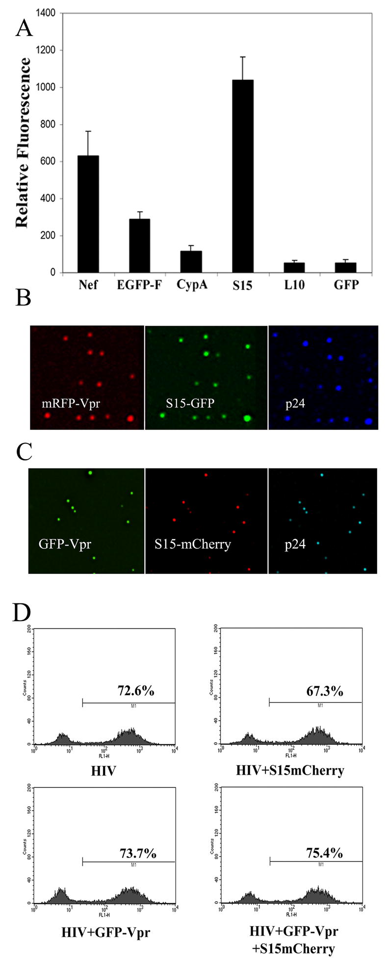
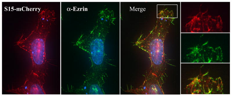
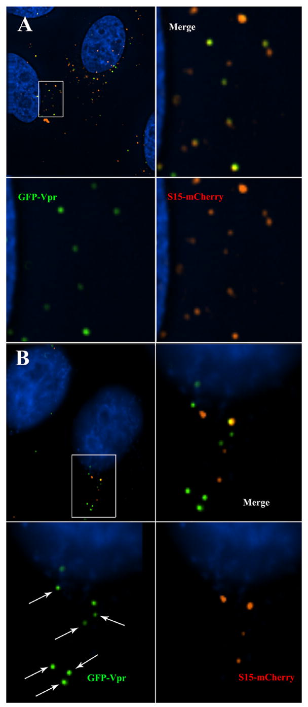
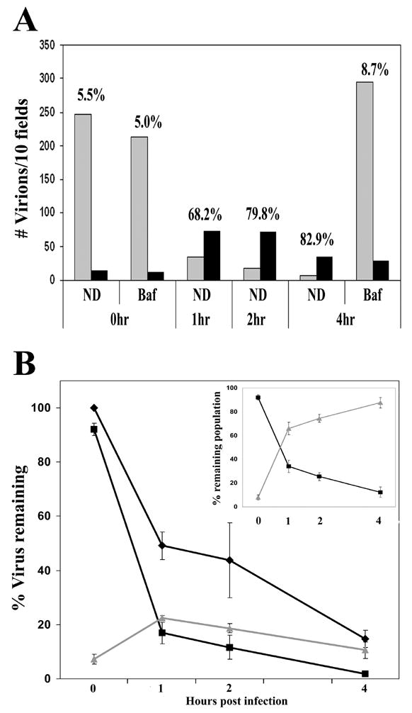
GFP-Vpr labeled HIV-1 virions have provided a method to visually examine the interactions between the virus and target cell during infection. However, existing methods to discriminate between virions that have been non-specifically endocytosed from those that have productively entered the host cell cytoplasm have remained problematic. Therefore, we examined the ability of a series of membrane-targeted fluorescent fusion protein constructs to be incorporated into virions. We find that a fluorescent protein fusion targeted to the plasma membrane by the addition of the N-terminal 15 amino acid sequence of c-Src (S15) is efficiently packaged into HIV virions. Using fluorescent proteins fused to this sequence, we have generated virions dually labeled with S15-mCherry and GFP-Vpr. Importantly, we can detect the loss of this S15-mCherry membrane signal following fusion. After infection with VSV-g pseudotyped HIV virions, we find a measurable, specific loss of membrane label during infection. This loss of fluorescence is not observed when fusion is prevented using bafilomycin A. This increased ability to discriminate between non-productively endocytosed virions and those actively undergoing steps in the infectious process will facilitate efforts to examine early steps in infection microscopically.


Figures

Figure 1
Figure 1. Incorporation of fluorescent fusion proteins into HIV-1 virions
A. mRFP-Vpr labeled virions produced from 293T cells cotransfected with the indicated construct were filtered through a 0.45 μm filter and bound to glass coverslips for 4 hours, fixed and immunostained with mAb specific for p24CA. GFP fluorescence of mRFP-Vpr positive virions was quantified using the data inspector function of Applied Precision software. Error bars represent the SEM of the GFP intensities measured in 20 or more mRFP-Vpr+ virions. Fusions containing the 15 N-terminal amino acids of c-Src were found to efficiently label HIV-1 virions whether an S15-GFP (B) or S15-mCherry (C) was used. (D) HeLa cells were infected with 40 ng VSV-g pseudotyped GFP reporter virus lacking fluorescent label or labeled with the indicated fluorescent proteins. GFP expression was measured 48 hours later by flow cytometry.
Figure 2
Figure 2. S15-mCherry localizes to dynamic areas of the plasma membrane in 293T cells
293T cells in a 6 well plate were transfected with 3 μg S15-mCherry using PEI. 24 hours post transfection, cells were stained with a monoclonal antibody specific for ezrin and analyzed by fluorescent microscopy. (INSERT) Membrane areas containing S15-GFP also stained positively for ezrin.
Figure 3
Figure 3. Virion incorporated S15-mCherry signal is lost following productive entry into target cells
HeLa cells were spinoculated (O’Doherty, Swiggard, and Malim, 2000) with VSV-g pseudotyped, S15-mCherry and GFP-Vpr labeled R7ΔEnv virions for 2 hours at 17º C. Virus was removed and replaced with 37ºC media and then additionally incubated for 2 hours at 37ºC in the presence (A) or absence (B) of BafA. Cells were then fixed, stained for DNA and imaged. Arrows indicate virions that do not have detectable S15-mCherry labeling.
Figure 4
Figure 4. Temporal analysis of S15-mCherry loss following infection
HeLa cells were spinoculated with VSV-g pseudotyped, S15-mCherry GFP-Vpr labeled R7ΔEnv virions for 2 hours at 17º C. Virus was removed and replaced with 37ºC media. Cells were incubated at 37ºC, fixed at the indicated time PI and imaged. GFP positive pucnta were than quantified and individually examined for the presence of above background mCherry signal. Panel A shows a representative experiment in which the number of virions observed per 10 Z-stack fields was calculated at the indicated time points in the presence or absence of BafA. Grey bars indicate the number of S15-mCherry positive virions and black bars indicate the number of S15-mCherry negative virions. Numbers above each time point indicate the percentage of GFP-Vpr positive virions lacking S15-mCherry signal. Panel B shows the total number of virions compiled from pooled data derived from 3 independent experiments. The input virion level was normalized to 100% in order to accommodate differences in initial observed viral input. The changes in the populations of total virions (◆) S15-mCherry positive virions (■) and S15-mCherry negative (▲) virions over time is shown. (Panel B inset) The relative amount of S15-mCherry positive virions (■) and S15-mCherry negative (▲) virions as a percentage of the remaining viral population. Error bars represent the standard error of the mean of 3 experiments.

References

    1. Bowman EJ, Siebers A, Altendorf K. Bafilomycins: a class of inhibitors of membrane ATPases from microorganisms, animal cells, and plant cells. Proc Natl Acad Sci U S A. 1988;85(21):7972–6. - PMC - PubMed
    1. Bretscher A, Edwards K, Fehon RG. ERM proteins and merlin: integrators at the cell cortex. Nat Rev Mol Cell Biol. 2002;3(8):586–99. - PubMed
    1. Bukovsky AA, Dorfman T, Weimann A, Gottlinger HG. Nef association with human immunodeficiency virus type 1 virions and cleavage by the viral protease. J Virol. 1997;71(2):1013–8. - PMC - PubMed
    1. Bukrinsky MI, Sharova N, Dempsey MP, Stanwick TL, Bukrinskaya AG, Haggerty S, Stevenson M. Active nuclear import of human immunodeficiency virus type 1 preintegration complexes. Proc Natl Acad Sci U S A. 1992;89(14):6580–4. - PMC - PubMed
    1. Campbell EM, Hope TJ. Gene therapy progress and prospects: viral trafficking during infection. Gene Ther. 2005;12(18):1353–9. - PubMed

Publication types

Substances